Pterygium surgery
Pterygium surgery is a medical procedure aimed at removing a pterygium, a benign growth of conjunctival tissue that forms on the surface of the eye and typically extends from the white of the eye (sclera) towards the cornea. Pterygia, sometimes called "surfer's eye," are often associated with prolonged exposure to UV rays, wind, dust, and dry environments. They can cause discomfort, redness, irritation, and even blurred vision if they encroach upon the cornea.

.png)
How does it work?
Learn more about the procedure / equipment

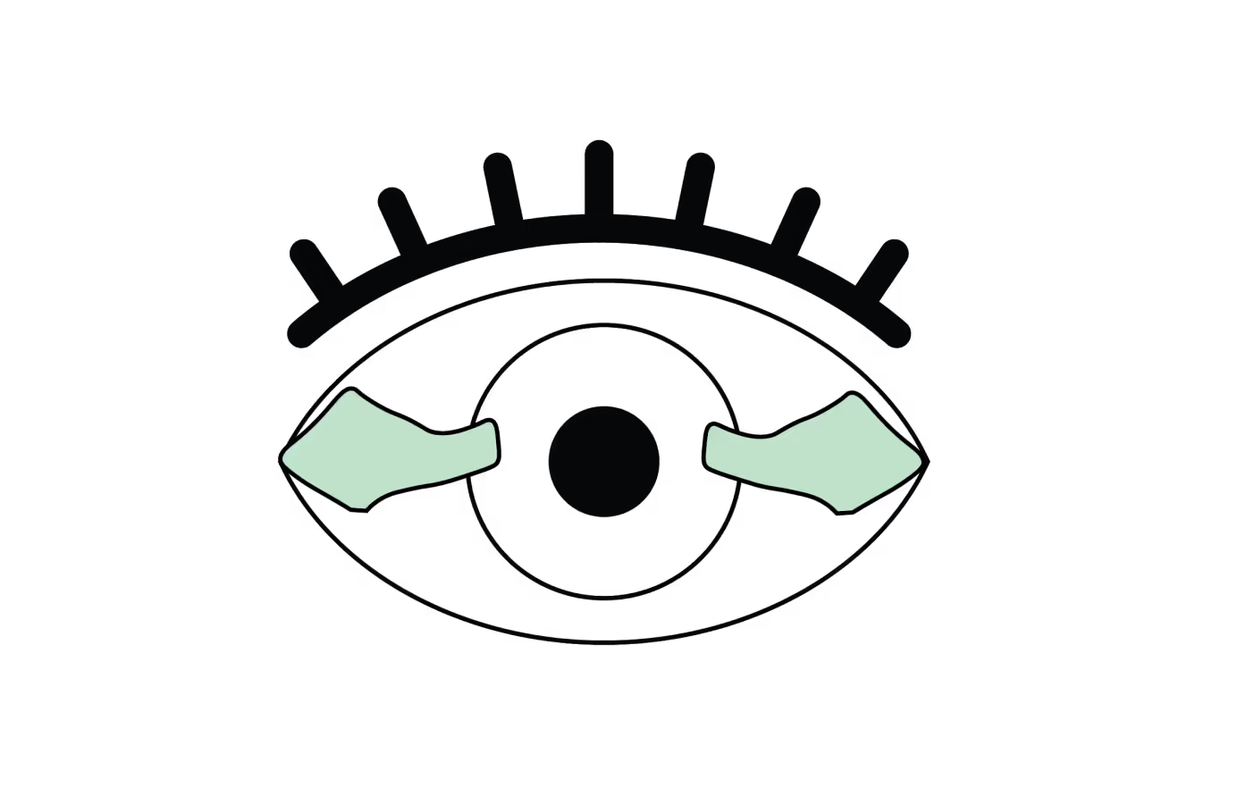
The pterygium is removed from the cornea and conjunctiva, leaving a visible conjunctival defect. Sometimes multiple pterygia are present simultaneously and can be excised during the same procedure.

A conjunctival graft or an amniotic membrane is placed over the bare area to promote healing and reduce the risk of recurrence.

The conjunctival autograft (or amniotic membrane) is integrated, the eye's appearance is restored, and symptoms such as irritation, redness, and blurred vision are improved or even resolved.
What is pterygium surgery?
Pterygium surgery is a procedure to remove a pterygium, a benign growth located on the conjunctiva (the transparent membrane covering the white part of the eye).
These growths are typically linked to excessive exposure to UV rays, dust, and dry environments. They can cause redness, irritation, and in advanced cases, lead to vision impairment if they invade the cornea.
The goal of the surgery is to relieve symptoms, improve vision, and restore the eye's appearance.
Why undergo pterygium surgery?
Pterygium surgery is generally recommended when the pterygium:
- Causes significant vision impairment by extending onto the cornea.
- Causes chronic eye irritation, redness, or discomfort not relieved by lubricating or anti-inflammatory eye drops.
- Presents an aesthetic concern for the patient.
What are the different techniques?
- Bare Sclera Technique: This is the simplest and oldest technique, where the pterygium is excised, leaving the sclera (the white part of the eye) exposed. This method has a high rate of pterygium recurrence and has largely been abandoned for this reason.
- Conjunctival Autograft Transplantation: In this technique, after the pterygium is removed, a piece of healthy conjunctival tissue is taken from another area of the eye and grafted to cover the treated area. This is the most common technique, as it significantly reduces the risk of recurrence.
- Amniotic Membrane Graft: An alternative to autograft when it is not possible, where an amniotic membrane (from a donor placenta) is used to cover the exposed area. It promotes healing and reduces the risk of recurrence.
- Fibrin Glue and Sutures: After pterygium excision, the conjunctival autograft (or amniotic membrane) can be secured using fibrin glue and/or sutures. Fibrin glue reduces surgical time and leads to less post-operative discomfort.
What are the benefits of this surgery?
- Symptom Relief: Pterygium can cause persistent irritation, redness, tearing, and a foreign body sensation. Surgery effectively relieves these symptoms, especially when other treatments like artificial tears and anti-inflammatory eye drops are insufficient.
- Improved Vision: When pterygium extends over the cornea, it can cause blurred vision or astigmatism by altering the cornea's shape. Its removal generally restores clear and stable vision, while preventing any further corneal deformation.
- Aesthetic Improvement: Pterygium can be very visible and cause aesthetic discomfort for some individuals. Surgery helps restore a more normal appearance to the eye, which can improve self-confidence and comfort in social interactions.
- Low Recurrence Rate with Modern Techniques: Techniques such as conjunctival autograft transplantation and amniotic membrane grafting have significantly reduced pterygium recurrence rates.
- Quick Procedure: Pterygium surgery is generally performed on an outpatient basis and lasts approximately 20-30 minutes. It is done under local anesthesia, making it a minimally invasive procedure.
- Reduced Discomfort with Advanced Techniques: The use of fibrin glue instead of traditional sutures to secure (auto-)grafts helps reduce postoperative discomfort, shorten the procedure duration, and accelerate healing.
What are the risks of surgery?
Although pterygium surgery is generally safe and effective, it carries certain potential risks and complications, like any ophthalmic procedure.
1. Pterygium Recurrence
- This is the main risk, with a higher rate in techniques without conjunctival autograft (or amniotic membrane).
- Prolonged UV exposure and an inflammatory predisposition increase the risk of pterygium regrowth.
2. Persistent Inflammation and Redness
- A postoperative inflammatory reaction is common and can last several weeks.
- It is usually well controlled with anti-inflammatory eye drops.
3. Infection
- Although rare, an infection can occur after surgery.
- Good eye hygiene and the use of antibiotic eye drops reduce this risk.
4. Dry eyes and discomfort
- Some patients experience persistent dryness after the procedure.
- Using artificial tears can help alleviate this discomfort, which is usually temporary.
5. Astigmatism or vision changes
- Pterygium excision can alter the curvature of the cornea and change astigmatism.
- In rare cases, additional optical correction may be necessary, but astigmatism generally tends to decrease rather than worsen after pterygium removal.
6. Graft dehiscence or displacement
- The conjunctival autograft (or amniotic membrane) can sometimes detach or shift.
- This may require further surgery to reposition or suture it.
Pterygium surgery is an effective procedure, but it is not without risks. Careful patient selection, an appropriate surgical technique, and rigorous postoperative follow-up help minimize complications and optimize results.
Sun protection and good ocular hydration are essential to prevent recurrence.
Les différentes types de greffes
Découvrez les kératoplasties (= greffes de cornée)
Frequently asked questions
If you have any further questions, please do not hesitate to contact us!
How effective is pterygium surgery?
Pterygium surgery is highly effective at providing lasting symptom relief and improving the eye's appearance. The use of advanced techniques, such as conjunctival autograft or amniotic membrane graft, significantly reduces the risk of recurrence and improves outcomes.
What are the causes of pterygium?
Pterygium is primarily associated with prolonged exposure to UV rays, dust, wind, and dry environments. Individuals who spend a lot of time outdoors without eye protection, such as surfers, farmers, and construction workers, are at higher risk. Genetics and certain climates also play a role.
Is a pterygium dangerous?
A pterygium is a benign (non-cancerous) growth and is generally not dangerous. However, if it extends onto the cornea, it can impair vision by causing astigmatism, scarring, or blurred vision. In such cases, its excision may be recommended.
Linked procedures
Discover other related surgical topics
Linked conditions
Book a consultation
Swiss Visio Montchoisi
1006 Lausanne, Switzerland