[DRAFT IA] Keratoconus symptoms: the 6 early signs to recognise
Keratoconus gradually distorts the cornea and blurs vision. Recognising its earliest signs means you can act early, before eyesight declines.

The earliest symptoms of keratoconus are blurred, distorted vision, an astigmatism that changes often and growing sensitivity to light. These signs usually appear in adolescence or young adulthood. Spotted early, they allow the cornea to be stabilised before vision declines for good. This article covers the 6 early signs, the risk factors, how the diagnosis is confirmed and when to seek advice.
What is keratoconus?
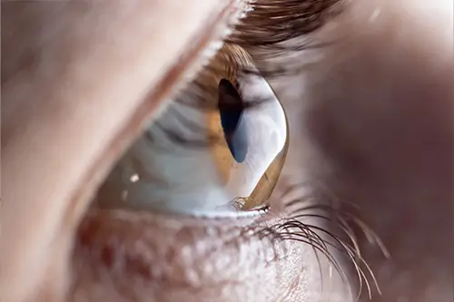
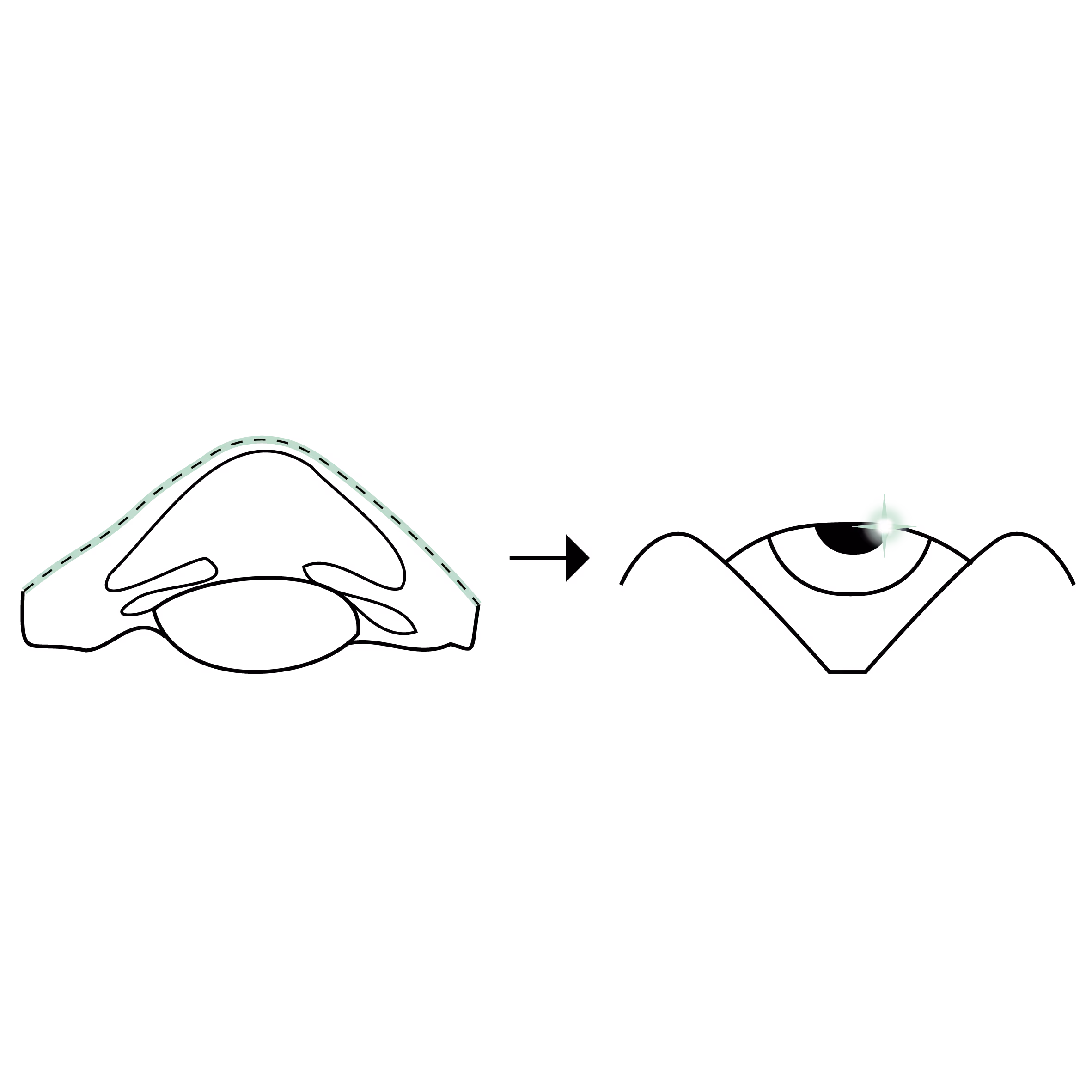
Keratoconus is a progressive disease of the cornea, the transparent lens at the front of the eye. The cornea gradually thins and bulges into a cone shape instead of staying evenly curved. This deformation disturbs how light reaches the retina and blurs vision.
The condition most often affects both eyes, sometimes asymmetrically. It frequently begins between the ages of 15 and 25, then tends to stabilise after 35 to 40. According to the European Society of Cataract and Refractive Surgeons (ESCRS), keratoconus affects roughly 1 person in 2,000 [VERIFIER_DONNEE: prevalence to be confirmed for the reference population].
The 6 early symptoms of keratoconus
Keratoconus progresses slowly and its first signs can be subtle. Here are the manifestations that should prompt attention.
1. Blurred, distorted vision
The corneal deformation creates an irregular astigmatism: straight lines look wavy, edges are poorly defined and images sometimes appear doubled in a single eye. This distorted vision is not fully corrected by standard glasses.
2. An astigmatism that changes often
A characteristic sign is the need to change your prescription frequently. If your glasses are adjusted several times in a short period, with a rising astigmatism, a corneal examination is warranted.
3. Increased sensitivity to light
Many patients describe glare, halos around light sources and marked discomfort at night, especially facing headlights. Night driving becomes uncomfortable.
4. Visual fatigue and headaches
The eye constantly compensates for the deformation, which leads to visual fatigue, a feeling of eye strain and sometimes headaches at the end of the day.
5. Declining vision despite glasses
When the astigmatism becomes too irregular, glasses are no longer enough. Switching to rigid contact lenses is often needed to restore sharp vision.
6. Itching and frequent eye rubbing
Repeated eye rubbing is both a risk factor and a warning sign. It is often linked to allergies or eczema. One rule matters: do not rub your eyes, as this accelerates the corneal deformation.
Who is affected and what are the risk factors?
Keratoconus results from a combination of predisposition and mechanical factors. The main risk factors are:
- chronic eye rubbing, a major aggravating factor
- ocular allergies and eczema, which encourage rubbing
- a family history of keratoconus
- certain genetic conditions, such as Down syndrome
- adolescence and young adulthood, the most active period of progression
Keratoconus is often accompanied by a progressive astigmatism. This is why any astigmatism that progresses rapidly in a young patient deserves a thorough corneal examination.
How is the diagnosis confirmed?
The diagnosis of keratoconus rests on a specialised examination of the corneal shape. Corneal topography maps the surface and thickness of the cornea and detects even very early deformations, before any loss of vision.
This is complemented by optical coherence tomography (OCT) of the cornea and a measurement of corneal thickness (pachymetry). These data confirm the diagnosis, assess the stage and track progression over time. These symptoms may suggest keratoconus, but only a specialist examination can establish the diagnosis. For a full assessment, you can consult Dr Arthur Hammer for keratoconus, an FMH ophthalmologist specialising in the cornea.
Why seeing a specialist early changes everything
Detecting keratoconus at an early stage is decisive. As long as the cornea keeps deforming, treatment can often slow its progression and preserve vision.
Corneal collagen cross-linking (CXL) is the reference treatment to stabilise progressive keratoconus. It strengthens the bonds between the cornea's collagen fibres to make it stiffer. The earlier it is performed, the better it preserves good vision. Other options, such as intracorneal ring segments, can improve the corneal shape at certain stages. The choice is made after a full assessment with a specialist in corneal surgery.
When should you seek urgent advice?
Some signs reflect rapid progression and warrant prompt consultation:
- a rapid drop in vision over a few weeks
- a sudden distortion of images
- a sharp pain with an abrupt loss of vision and a red eye, which may suggest corneal hydrops, a rare complication
In these situations, a specialist ophthalmic opinion is needed quickly.
FAQ: common questions about keratoconus symptoms
At what age does keratoconus appear?
Keratoconus most often begins in adolescence or young adulthood, roughly between 15 and 25. This is when the disease progresses fastest. It then tends to stabilise around 35 to 40. An early diagnosis allows progression to be monitored and treatment to be timed correctly to preserve vision.
Does keratoconus cause blindness?
Keratoconus does not cause blindness. It can, however, lead to a significant loss of vision if left untreated. In the vast majority of cases, vision is preserved with contact lenses, cross-linking or, at advanced stages, a corneal transplant. Only a specialist examination can assess the stage and the appropriate treatment.
Can keratoconus be corrected with glasses?
In the early stages, glasses may be enough to correct the astigmatism. When the deformation becomes too irregular, glasses no longer correct vision properly. Rigid or scleral contact lenses then become necessary to restore sharp vision.
Can eye rubbing cause keratoconus?
Chronic eye rubbing is a recognised risk factor for keratoconus and its progression. It applies repeated mechanical stress to a weakened cornea. This is why ophthalmologists recommend avoiding eye rubbing and treating the ocular allergies that encourage it.
Is keratoconus hereditary?
There is a familial component: having an affected parent increases the risk. Keratoconus is not strictly a hereditary disease, however, as mechanical factors such as eye rubbing also play a role. Screening with corneal topography is advised for the relatives of an affected patient, especially during adolescence.
How do I know whether my astigmatism is hiding keratoconus?
An astigmatism that rises rapidly, forces frequent changes of glasses or is not fully correctable should raise the suspicion of keratoconus, especially in a young patient. Only corneal topography performed by an ophthalmologist can tell the difference and establish a reliable diagnosis.
In summary
Keratoconus is recognised by distorted vision, a progressive astigmatism and light sensitivity, especially in adolescents and young adults. Spotting these signs early allows the cornea to be stabilised and vision preserved over the long term. If you recognise several of these symptoms, book an appointment with Dr Arthur Hammer, an FMH ophthalmologist specialising in the cornea in Lausanne and Geneva, for a corneal topography assessment.
Les motifs de consultations liés
.avif)
.avif)
Articles relatifs
Découvrez nos autres articles qui pourrait vous intéresser.
Book a consultation
Swiss Visio Montchoisi
1006 Lausanne, Switzerland