Auto-slet: Limbal Stem Cell Transplant
Auto-SLET (Simple Limbal Epithelial Transplantation) helps to repair the surface of the eye when it has been severely damaged. It is an autologous transplant (meaning the tissue is harvested from the patient themselves), in which a small piece of limbal tissue is taken from the healthy eye and placed onto the affected eye to facilitate healing. This procedure often results in improved vision and a significant reduction in ocular pain.

How does it work?
Learn more about the procedure / equipment
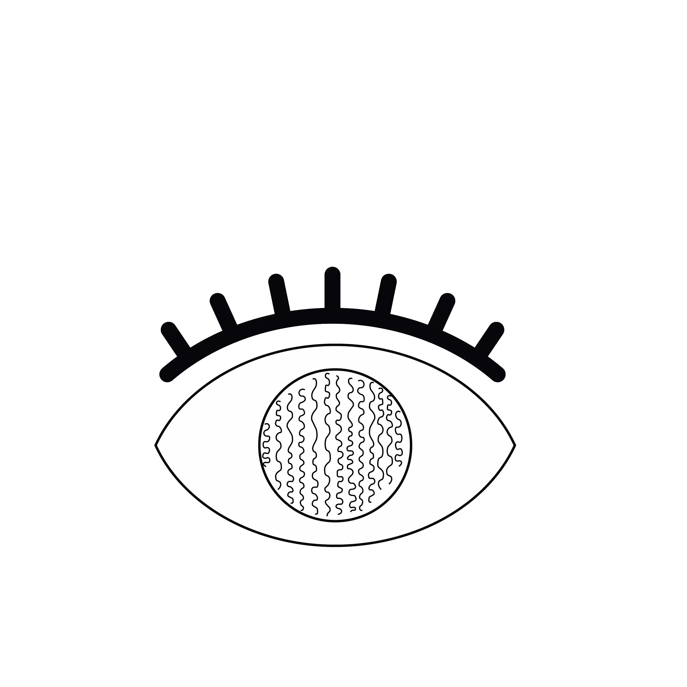
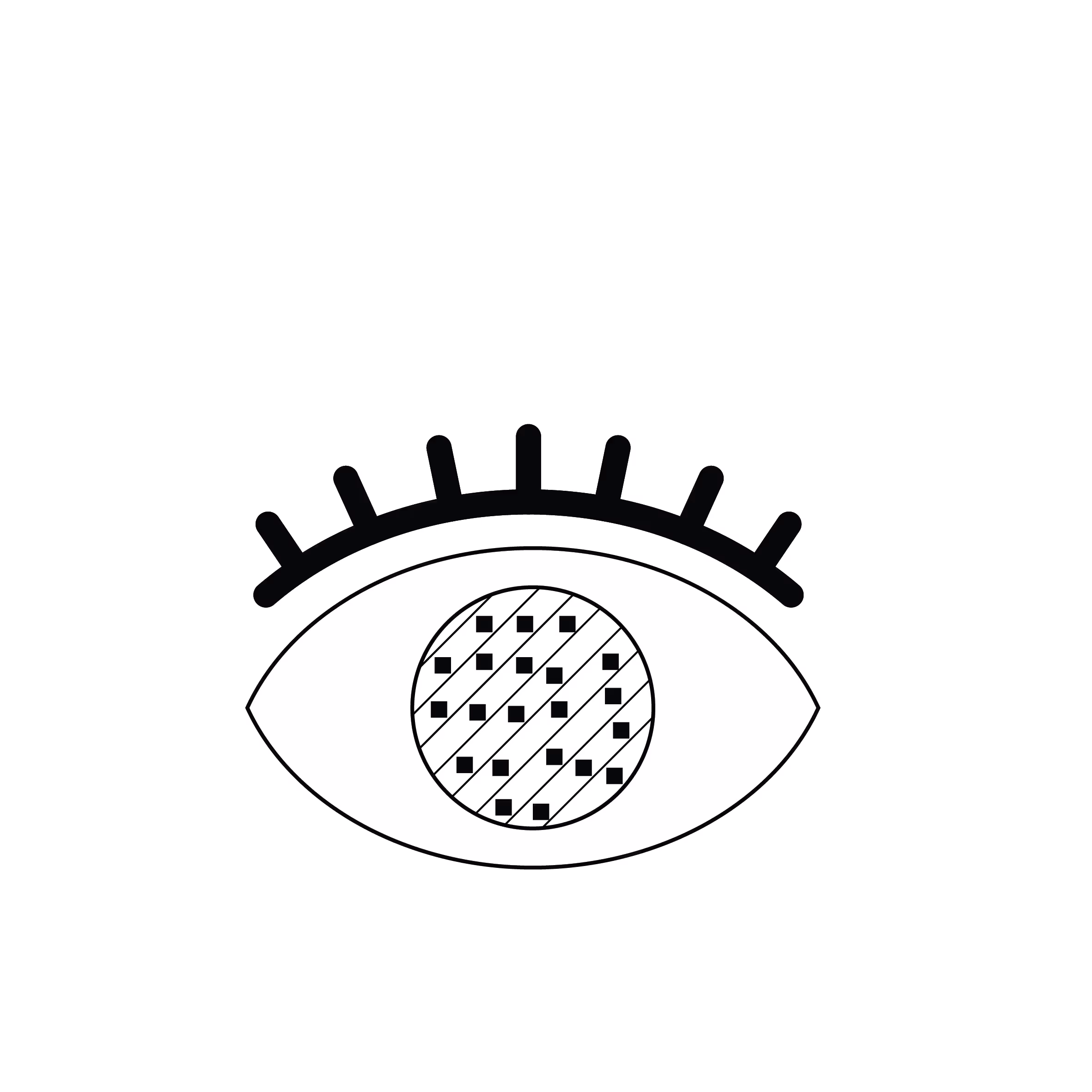
1. Recipient eye with scarred surface
The diseased eye presents an opaque, irregular, and often vascularized corneal surface, typical of a limbal stem cell deficiency, often caused by trauma or a chemical burn.
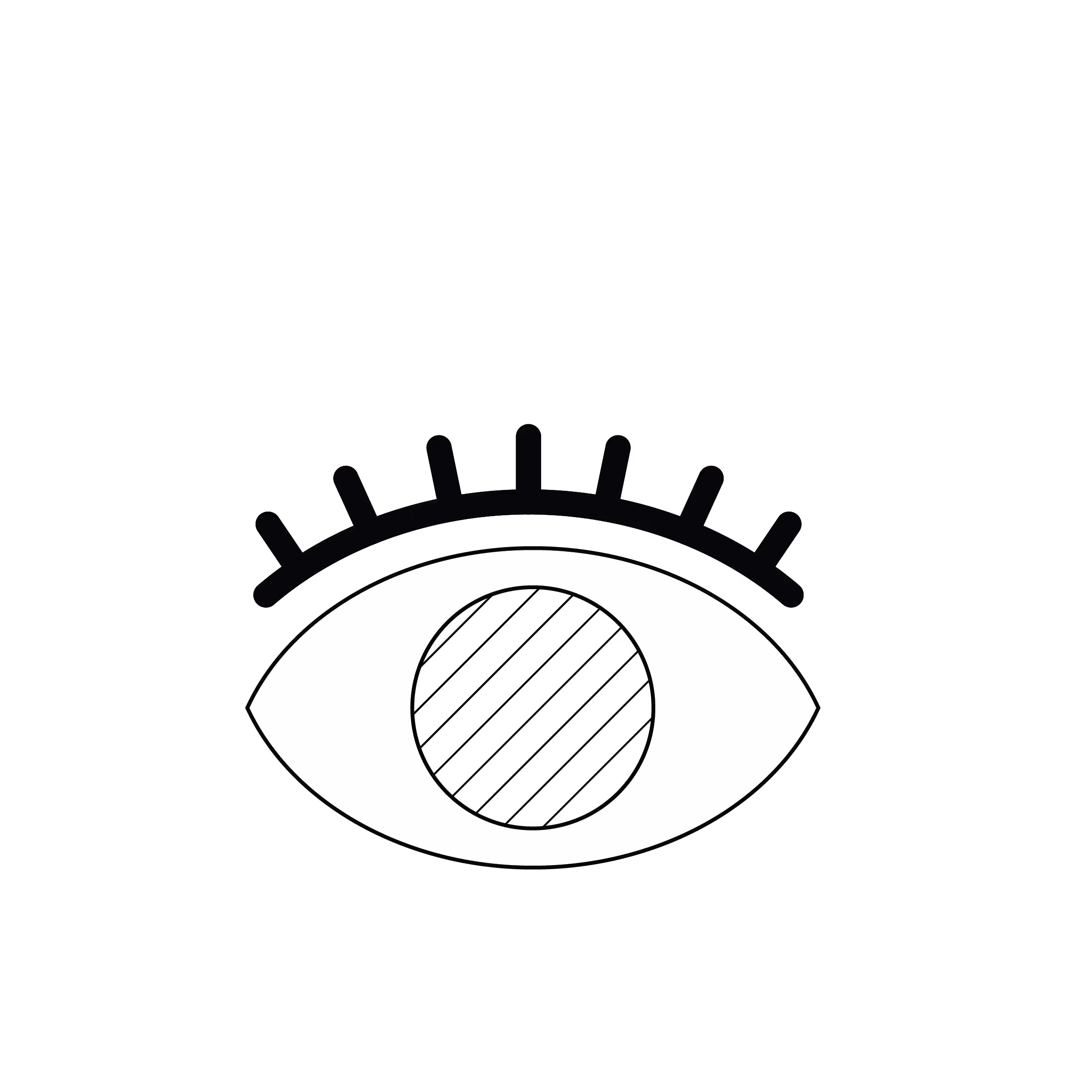
2. Ocular Surface Preparation
The surgeon carefully removes the abnormal and scarred tissue from the corneal surface of the recipient eye, exposing the underlying corneal stroma to create a clean bed for the graft.
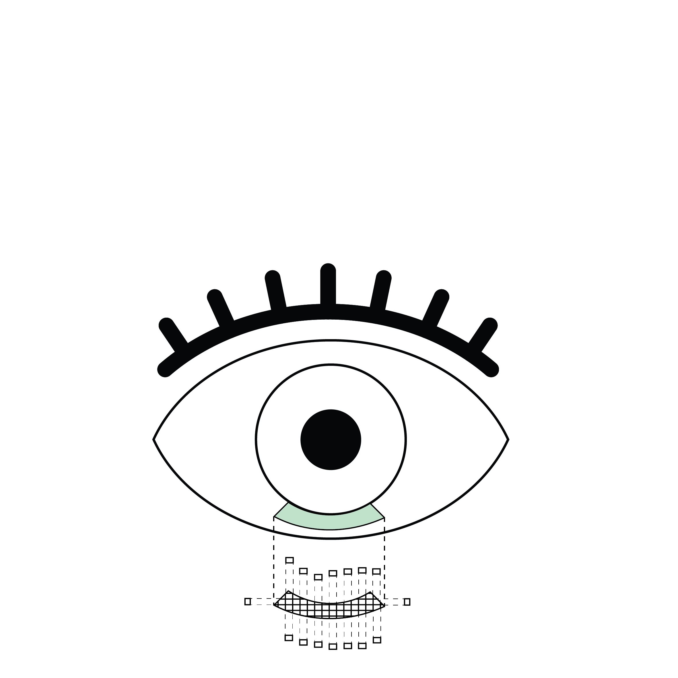
3. Harvesting limbal tissue from the healthy eye
A thin strip of healthy corneal limbal tissue (approximately 3 mm × 1 mm) is harvested from the healthy/donor eye and cut into small fragments (approximately 16 small squares). This tissue contains essential stem cells for the renewal of the corneal surface epithelium.
4. Fragment Transplantation onto the Recipient Eye
The small fragments of limbal tissue transplanted are spread over an amniotic membrane placed on the cornea of the diseased/recipient eye. The fragments are secured with biological glue and covered by a therapeutic contact lens to protect them.

5. Proliferation and Migration of Epithelial Cells
The limbal stem cells derived from the fragments begin to multiply and migrate to gradually cover the cornea, restoring a healthy and functional epithelium.
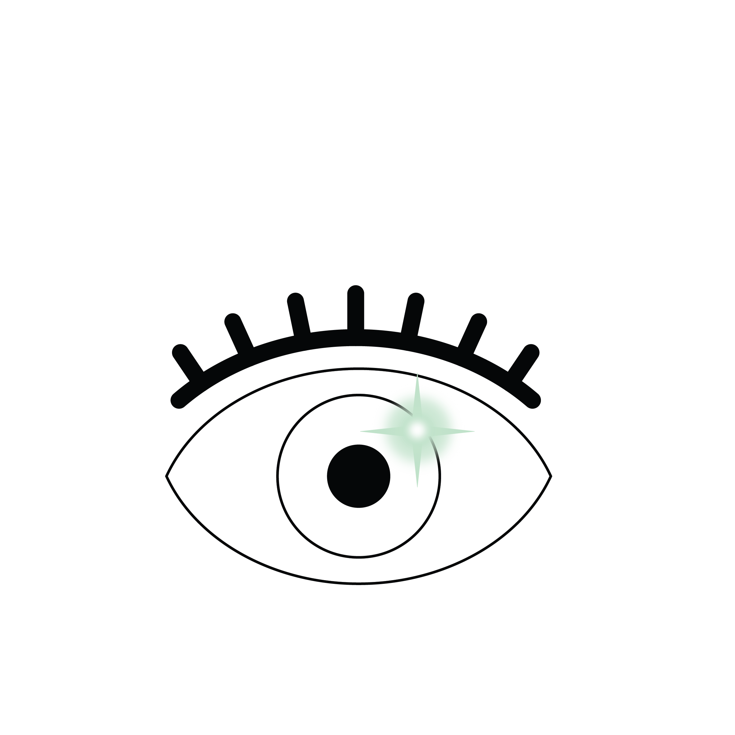
6. Ocular Surface Restoration
After several weeks, the corneal surface is clear and regular, with improved vision and a significant reduction in symptoms. The graft is stable and integrated.
What is Auto-SLET?
Auto-SLET (Simple Limbal Epithelial Transplantation) is a surgical technique used to treat patients with unilateral limbal stem cell deficiency (affecting only one eye). It involves harvesting a small quantity of stem cells from the limbus, the transition zone between the cornea and the conjunctiva, of the patient's healthy eye and transplanting them into the damaged eye.
Unlike a full graft or an allogeneic transplant (using a donor), Auto-SLET uses the patient’s own cells (autologous), meaning there is no risk of immune rejection. This eliminates the need for long-term immunosuppressive therapy. Once the ocular surface has been restored, if the cornea remains scarred, it may be possible to perform a full-thickness corneal transplant (PKP) or an anterior lamellar graft (DALK or SALK) to clear the vision further.
What is the procedure?
- Preparing the receiving eye: The surgeon removes scar tissue or abnormal growths from the corneal surface of the damaged eye to create a clean "bed" for the transplant.
- Limbal sampling: A tiny segment (approximately 3 mm × 1 mm) of limbus is harvested from the healthy eye. This sample is usually taken from the upper or lower limbus, as these areas are protected by the eyelid and have less impact on the eye's blood supply.
- Fragmentation and placement: The harvested tissue is cut into 12–16 small fragments and arranged onto an amniotic membrane placed over the damaged cornea. These fragments act as the source of new stem cells.
- Fixation: The fragments and membrane are secured using biological glue, and a therapeutic bandage contact lens is applied for protection.
- Healing: Over several weeks, the stem cells proliferate from the fragments to recolonise and restore the corneal surface.
What are the risks and benefits?
Benefits
- Long-lasting restoration of the ocular surface.
- Improvement in vision, comfort and corneal transparency.
- Minimally invasive, requiring only a very small donor sample.
- No immunosuppression required, as the cells are the patient's own.
- Suitable for both children and adults.
Risks
- Transplant failure if the fragments do not integrate correctly.
- Incomplete healing or a recurrence of neovascularisation (abnormal blood vessel growth).
- Complications related to the amniotic membrane (e.g., displacement or tearing).
- A theoretical (though rare) risk of stem cell deficiency in the healthy donor eye.
- Standard postoperative risks such as infection or inflammation.
Indications and Contraindications
Auto-SLET is primarily indicated for unilateral (one-sided) deficiency caused by:
- Chemical or thermal burns.
- Severe corneal or conjunctival injuries.
- Corneal neovascularisation due to serious infections or localised autoimmune conditions.
- Failure of previous ocular surface reconstructions.
It is not suitable for bilateral cases (both eyes affected). In such instances, an Allo-SLET (using donor tissue) or CLET (cultivated limbal epithelial transplantation) would be preferred.
Absolute Contraindications
- Bilateral deficiency: If both eyes are affected, healthy tissue cannot be harvested.
- Diseased donor eye: Severe dryness, chronic conjunctivitis or keratopathy in the "healthy" eye makes sampling unsafe.
- Active infection: In either the recipient or donor eye.
- Non-compliance: Success depends on rigorous follow-up and adherence to postoperative eye drops.
Relative Contraindications
- Uncontrolled inflammation: Active autoimmune flare-ups increase the risk of failure.
- Severe dry eye: Without an adequate tear film, the graft may struggle to survive.
- Eyelid abnormalities: Conditions such as entropion (inward-turning eyelid), trichiasis (misdirected eyelashes) or blinking disorders.
Les différentes types de greffes
Découvrez les kératoplasties (= greffes de cornée)
Frequently asked questions
If you have any further questions, please do not hesitate to contact us!


Linked conditions
Book a consultation
Swiss Visio Montchoisi
1006 Lausanne, Switzerland