DMEK corneal transplant: understanding the procedure and recovery for Fuchs dystrophy
The DMEK corneal transplant replaces only the thin, damaged endothelial layer of your cornea. Here is how the procedure is carried out and how recovery unfolds.
Created on
20.07.2026
·
Updated on
10.08.2026

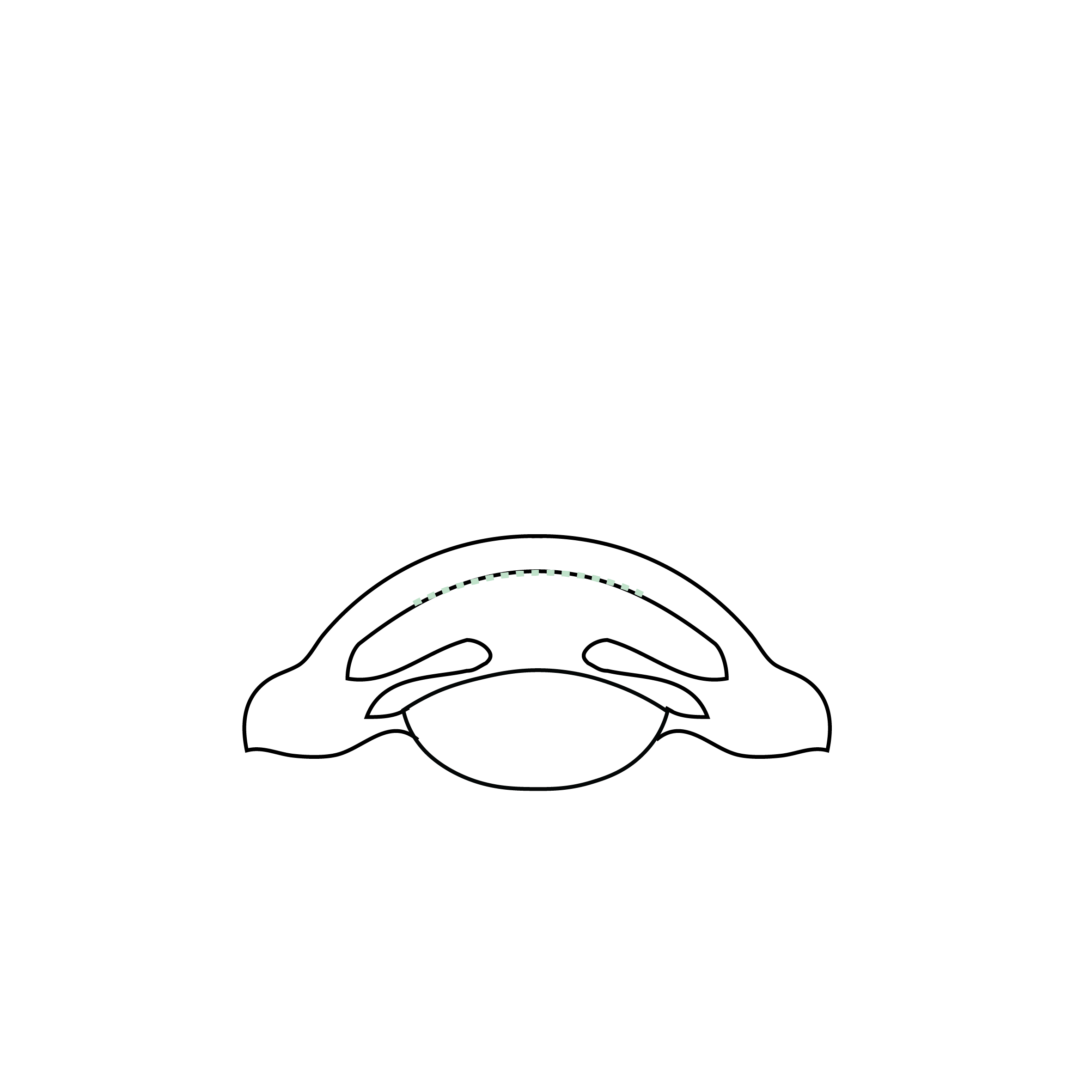
The DMEK corneal transplant involves replacing only the thin inner layer of your cornea, the endothelium, with healthy donor tissue. It is mainly intended for patients with Fuchs dystrophy or corneal oedema. The procedure is generally performed on an inpatient basis, under local anaesthetic, and generally takes less than an hour. Being told you need a transplant can be worrying. This page explains, step by step, what DMEK is, who it is offered to, how the operation is carried out, recovery day by day, the expected results, the risks and reimbursement in Switzerland.
DMEK, a transplant that replaces only the inner layer of the cornea
DMEK (Descemet Membrane Endothelial Keratoplasty) is a lamellar transplant technique that replaces only Descemet's membrane and the endothelium, the deepest layer of the cornea. It leaves the rest of your cornea in place, unlike a penetrating transplant that replaces the full thickness.
The corneal endothelium is a layer of cells that pumps water out of the cornea. When these cells no longer function, the cornea becomes waterlogged, turns cloudy and vision declines.
DMEK aims to restore this pump function by providing healthy endothelial cells. The transplanted graft is extremely thin, in the order of 15 to 20 microns thick or so. This thinness explains the visual quality that is often achieved.
This approach became popular in the 2000s and is now the reference for isolated endothelial disorders. Dr Hammer, an FMH consultant ophthalmic surgeon specialising in the cornea, performs it in French-speaking Switzerland. You will find more technical detail on the page dedicated to DMEK, ultra-thin endothelial lamellar keratoplasty.
DMEK, DSAEK and penetrating transplant: what are the differences
DSAEK is another endothelial transplant, but the graft is thicker because it includes a thin layer of stroma. DMEK, being thinner, often offers faster visual recovery and a lower risk of rejection, according to studies.
The penetrating transplant, or penetrating keratoplasty, replaces the full thickness of the cornea. It remains useful when the cornea is affected in depth. The choice between these techniques depends on your condition. For a complete overview, see the page on corneal transplants and keratoplasties.
Who is DMEK indicated for
DMEK is indicated when the corneal endothelium is failing while the rest of the cornea is healthy. The aim is to treat the cause of the oedema without touching the superficial layers.
The first indication is Fuchs dystrophy, a common hereditary disease that gradually wears down the endothelial cells. It often shows as blurred vision in the morning that improves during the day.
DMEK also applies to endothelial decompensation after eye surgery, for example after cataract surgery. This is referred to as secondary corneal oedema.
The main situations where an endothelial transplant may be considered are the following:
Not all eyes are suited to DMEK. Certain particular anatomies point towards DSAEK. Only a specialist examination can establish the diagnosis and select the appropriate technique. You can find out more about the different corneal transplants before your consultation.
The DMEK procedure, step by step
The DMEK procedure is a microsurgical operation carried out on an inpatient basis, most often under local anaesthetic.
The preoperative assessment
Before the operation, a full assessment is carried out. It includes a measurement of corneal thickness by pachymetry, an analysis of the endothelial cells by specular microscopy and imaging by corneal and retinal OCT where the clarity of the optical media allows.
This assessment confirms the indication and rules out other causes of reduced vision, such as a retinal disorder. Dr Hammer then explains the benefit-risk balance and answers your questions.
The surgical technique
The surgeon first removes the diseased Descemet's membrane through a small incision. He then prepares the thin graft obtained from a tissue bank, and gently introduces it into the eye.
The graft is unfolded carefully, then pressed against the cornea using an air bubble or gas injected into the eye. This bubble acts to hold it in place during the first few hours to days.
The procedure generally lasts around 30 to 60 minutes, depending on the case. The incisions are so small that they usually require no sutures, or very few.
Just after the operation
You remain lying down for a while in the recovery room, on your back, with your face turned towards the ceiling. This position helps the bubble to keep the graft in place.
DMEK causes little pain. You may feel some discomfort, a gritty sensation or watering for one to two days. Treatment with eye drops is started.
Recovery after DMEK, day by day
Recovery after DMEK rests on a simple rule: keep your head facing up to let the bubble press the graft into place. This positioning determines the proper adhesion of the grafted tissue.
During the first few hours to days, the surgeon asks you to stay lying on your back as much as possible, with your face turned towards the ceiling and, above all, to avoid turning your face towards the floor (looking down). This face-up positioning instruction is essential, including at night.
Vision is very blurred at first, because of the air bubble and the residual oedema. This is normal. The bubble is gradually absorbed over a few days to a week.
Here are the usual milestones of recovery, as a guide:
The eye drops, in particular corticosteroids and/or calcineurin inhibitors, are continued for several months to limit the risk of rejection. You must avoid rubbing the operated eye during the first week and must follow the drop schedule scrupulously.
Time off work from a few days to two weeks is common, depending on your occupation. Dr Hammer adapts these instructions to your situation during follow-up.
Results and visual recovery times
DMEK aims to restore the cornea's transparency and thereby to improve vision. Results are gradual and depend on the initial state of your eye.
Many patients notice an initial improvement within the first few days to weeks. Vision then continues to sharpen over several months, as the oedema clears completely.
DMEK is recognised for the quality of vision it can provide, often better than that of thicker transplants, according to data from the literature. The result also depends on any associated conditions, such as a retinal disorder.
No surgeon can guarantee a precise result. The aim is to improve your vision and comfort, while respecting the particular features of your eye.
Possible risks and complications of DMEK
DMEK is a well-established surgery, but like any procedure it carries risks that transparency requires you to be aware of. They remain limited in most cases.
The most frequent complication is partial detachment of the graft. The grafted tissue may not adhere completely. An air reinjection, known as re-bubbling, is then carried out, a simple and quick procedure.
The reported re-bubbling rate varies between studies, generally between 10 and 20%. In experienced hands, it can be reduced to less than 5%, as is the case for Dr Hammer. This additional step does not compromise the final result.
Graft rejection is possible, but rarer with DMEK than with DSAEK and penetrating transplants, according to the literature. The prolonged anti-inflammatory drops serve precisely to reduce this risk.
Other complications may occur, such as raised eye pressure, an infection or graft failure requiring a further operation. These situations remain uncommon.
Regular follow-up allows these problems to be detected early and acted on quickly. This is why postoperative check-ups must never be neglected.
When to seek urgent care after DMEK
An urgent consultation is necessary as soon as signs suggesting rejection or a complication appear. Spotting these signs early helps to preserve the graft.
You should quickly contact your surgeon or an ophthalmology department in the event of:
- Acute or increasing eye pain: sharp, throbbing pain not controlled by the usual painkillers (suggesting raised eye pressure, a pupillary block from the air or gas bubble, or an infection).
- Sudden or marked drop in vision: a clear fall in visual acuity compared with the initial postoperative improvement, or a sudden persistent blur.
- Marked or worsening eye redness: particularly if accompanied by eyelid swelling.
- Purulent discharge or abnormal watering: yellowish or greenish, suggesting endophthalmitis or infectious keratitis.
- Intolerable light sensitivity: recent-onset extreme sensitivity to light.
- Nausea or vomiting associated with eye pain: a classic sign of a severe rise in eye pressure (acute glaucoma).
These symptoms do not always mean a serious complication. But only a specialist examination can tell them apart and adjust treatment without delay.
In practice at Dr Hammer's practice
Dr Arthur Hammer, an FMH consultant ophthalmic surgeon specialising in the cornea, carries out the full preoperative assessment and generally operates on an inpatient basis, in particular at the Montchoisi clinic in Lausanne. Each DMEK indication is established after a detailed examination of your cornea and endothelium. You receive written postoperative precautions, positioning instructions and a precise eye-drop schedule. Close follow-up is arranged in the days and then the weeks after the procedure, in order to check the proper adhesion of the graft and adjust your treatment.
Cost and reimbursement of DMEK by Swiss basic health insurance (LAMal)
The DMEK corneal transplant is a medically indicated procedure, covered by Swiss basic health insurance (LAMal) when the medical criteria are met. It is not a cosmetic procedure.
In practical terms, LAMal covers the procedure, the graft and the necessary medical follow-up. Your contribution is in principle limited to your annual deductible and the 10% co-payment, within the legal limits.
Before the procedure, a guarantee of coverage can be requested from your insurer. The practice's secretariat will assist you with these administrative steps. For any question about your situation, please get in touch with your insurer.
For patients not covered by Swiss health insurance, a quote is provided before the procedure.
FAQ: your questions about the DMEK corneal transplant
Is the DMEK transplant painful?
The procedure itself is not painful. It is performed under local anaesthetic, often combined with light sedation to help you relax, so you do not feel the operation. In the days that follow, you may feel some discomfort, a gritty sensation or slight watering. These sensations are usual and settle quickly. Eye drops and, if needed, a simple painkiller are most often enough to keep you comfortable. Genuine pain is unusual and should prompt you to contact your surgeon.
How long does it take to recover vision after DMEK?
Visual recovery is gradual. Vision remains blurred in the first few days because of the air bubble and the oedema. It then clears over the following weeks. In most cases, useful vision is regained between a few days and a few weeks, then stabilises over a few months. The timescale varies with the initial state of your eye and any associated conditions. Regular follow-up allows this progress to be tracked step by step.
Why must I keep my head facing up after the operation?
During DMEK, the graft is pressed against your cornea by an air or gas bubble. This bubble naturally rises upwards. By keeping your face turned towards the ceiling, you help it to hold the graft firmly in place until it adheres. This positioning, strict in the first few hours after the operation, is decisive for the success of the transplant. Your surgeon will tell you the exact duration and the times when you can get up.
What is re-bubbling?
Re-bubbling is a reinjection of air or gas into the eye. It is used when the graft has not stuck perfectly to the cornea and partly detaches. This simple, quick procedure is carried out to press the graft back into place. It is needed in a proportion of cases, without compromising the final result in most situations. It is therefore not a failure, but a sometimes useful step in the care pathway.
What is the risk of rejection after DMEK?
Graft rejection does occur, but it is rarer with DMEK than with other types of transplant such as DSAEK and penetrating keratoplasty, according to studies. The very thin graft and the small amount of transplanted tissue explain this reduced risk. The anti-inflammatory drops prescribed for several months serve to limit it further. If you have reduced vision, or a red or painful eye, seek advice without delay. Only a specialist examination can confirm rejection and treat it quickly to preserve the graft.
Is DMEK reimbursed by LAMal?
Yes, the DMEK corneal transplant is covered by Swiss basic health insurance (LAMal) when the medical indication is established, for example symptomatic Fuchs dystrophy or corneal oedema. It is not a cosmetic procedure. Your contribution is in principle limited to your annual deductible and the 10% co-payment, according to the rules in force. A guarantee of coverage can be requested from your insurer. The practice's secretariat will help you with these steps.
Can both eyes be operated on at the same time?
Generally, both eyes are not operated on the same day. The surgeon treats one eye first, checks the graft has taken and that recovery is under way, then plans the second eye later. This interval helps to make each stage safe and to organise the postoperative positioning. The strategy depends on your visual difficulty and the extent of involvement of each eye. Dr Hammer will define with you the schedule best suited to your situation.
Which activities should be avoided after a DMEK transplant?
After DMEK, you must avoid rubbing the operated eye, as well as swimming pools, saunas and dusty environments during the stated period. Intense physical effort and carrying heavy loads are limited at first. Face-up positioning takes priority during the first few hours to days. You then gradually resume your activities according to your surgeon's instructions. As every situation is different, follow precisely the recommendations given to you at follow-up.
In conclusion
The DMEK corneal transplant makes it possible to treat Fuchs dystrophy and corneal oedema by replacing only the damaged endothelial layer. In most cases it offers good-quality visual recovery and a reduced risk of rejection, at the price of strict positioning during the first few days.
Every eye is different. Only a specialist examination can confirm the indication and choose the most suitable technique. To assess your situation, you can request a consultation with Dr Arthur Hammer, FMH consultant ophthalmic surgeon, or book an appointment directly from the practice's home page.



Related articles
Discover our other articles that might interest you

Book a consultation
Swiss Visio Montchoisi
1006 Lausanne, Switzerland