Fuchs Dystrophy: Symptoms, Progression and Corneal Treatments
Fuchs dystrophy slowly damages the corneal endothelium. Symptoms, stages, diagnosis and treatments explained by Dr Hammer.
Created on
20.07.2026
·
Updated on
08.08.2026
Fuchs dystrophy is a genetic corneal condition that progressively damages the endothelium, the inner layer responsible for keeping the cornea clear. It causes blurred vision, often more marked in the morning, followed by halos and increasing visual discomfort. This slow progression can be worrying, but solutions exist at every stage. In this article, Dr Arthur Hammer, FMH consultant ophthalmic surgeon in Lausanne, explains the symptoms, the progression by stages, the diagnosis and the current treatments for Fuchs dystrophy, from eye drops to endothelial keratoplasty.
What is Fuchs dystrophy?
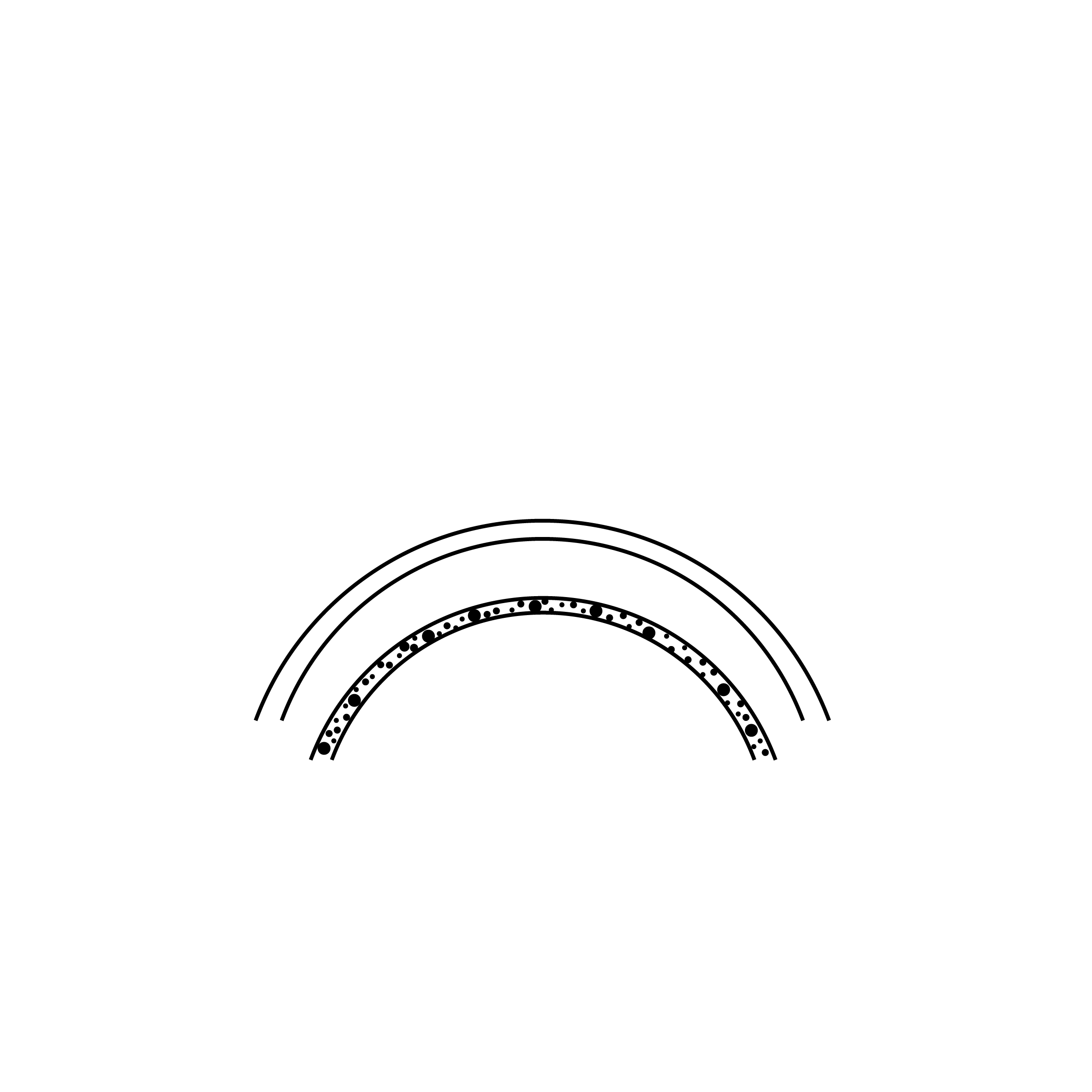
Fuchs dystrophy is a chronic corneal condition that affects the corneal endothelium, a thin layer of cells lining the inner surface of the cornea. These cells act as a pump.
Their role is to continuously remove the water contained within the cornea. This work keeps the cornea dehydrated, and therefore perfectly clear, an essential condition for sharp vision.
In Fuchs dystrophy, these endothelial cells decline more quickly than normal. The cornea then retains water, swells and loses its clarity, which is known as corneal oedema.
A characteristic sign appears early: cornea guttata. These are small outgrowths, the guttae, that form on Descemet membrane, just beneath the endothelium.
At birth, corneal endothelial cell density ranges between approximately 4'000 and 6'000 cells per mm². A natural loss of around 0.6% per year is then observed, leaving the adult endothelium with an average of approximately 2,000 to 3,000 cells per mm². These cells do not regenerate, which accounts for the progressive and irreversible nature of their loss.
What are the causes and risk factors?
Fuchs dystrophy is a condition that is mainly genetic in origin, often inherited in an autosomal dominant pattern. An affected parent can therefore pass the predisposition on to their children.
Several genes have been identified in recent studies, in particular variations in the TCF4 gene. The condition nonetheless remains multifactorial and its expression varies from one person to another.
Age is the main factor in progression. Troublesome symptoms most often appear after the age of 50, even though the first signs may be visible earlier on examination.
A female predominance is described: the condition is thought to affect women more than men.
Certain situations can accelerate decompensation, such as cataract surgery on a cornea that is already fragile. A careful preoperative assessment helps anticipate this risk.
What are the symptoms and how does the condition progress?
The symptoms of Fuchs dystrophy result from the build-up of water in the cornea, which blurs vision and progresses in stages over several years. The most typical sign is blurred vision on waking.
During the night, the closed eyelids limit evaporation, so the cornea becomes saturated with water. Vision then clears during the morning as the cornea dehydrates.
Over time, other problems appear: halos around lights, glare, reduced contrast and increased sensitivity to light. These signs reflect more pronounced oedema.
At advanced stages, the oedema can reach the surface of the cornea and cause painful blisters, known as bullous keratopathy. Pain occurs when these blisters burst.
Progression remains slow and variable. Several stages can be broadly distinguished, summarised below, which help Dr Hammer tailor monitoring and treatment.
These symptoms may suggest Fuchs dystrophy, but also other corneal conditions. Only a specialist examination can make the diagnosis with certainty.
How is the diagnosis made?
The diagnosis of Fuchs dystrophy is based on a complete ophthalmic examination, focused on the cornea and its endothelium. The first step is examination with the slit lamp.
This microscope allows Dr Hammer to directly identify the guttae and any oedema. He also assesses the general appearance of the cornea and the other structures of the eye.
Specular microscopy or confocal microscopy completes the examination. It counts the endothelial cells and measures their density, key information for monitoring progression.
Pachymetry measures the thickness of the cornea. A thickened cornea indicates water retention and helps quantify the oedema over successive consultations.
Finally, corneal OCT provides a precise cross-section of the different layers. This imaging examination refines the assessment and guides the treatment decision, especially before any potential procedure.
What are the treatments according to the stage?
The treatment of Fuchs dystrophy depends on the stage of the condition and the discomfort experienced, ranging from eye drops to corneal grafting. In the early stages, the aim is to limit the oedema.
Hypertonic sodium chloride eye drops help to draw out some of the water at the surface. Some patients also find morning relief by gently drying the cornea with warm air.
These measures do not cure the condition, but they improve visual comfort in most mild cases. They often make it possible to postpone the need for surgery.
When the oedema becomes permanent and interferes with daily life, endothelial keratoplasty becomes the reference option. It involves replacing only the diseased inner layer of the cornea.
Two main techniques exist. DMEK, a thin endothelial lamellar keratoplasty, grafts a very thin membrane and offers rapid visual recovery in many cases.
The DSAEK technique grafts a slightly thicker lamella. The choice between these two options is a matter for discussion between the patient and the surgeon, depending on the state of the eye.
These procedures are among the corneal grafts, or keratoplasties. Endothelial keratoplasty is now becoming the most common corneal graft procedure.
In certain selected cases, an approach without grafting is possible: DWEK/DSO. It involves removing a small central area of diseased membrane to allow the healthy peripheral cells to repopulate the cornea.
This option remains reserved for specific situations, with a good-quality peripheral endothelium. Only a specialist examination can determine whether it is feasible in your case.
In practice at Dr Hammer's clinic
Dr Arthur Hammer, FMH consultant ophthalmic surgeon and corneal specialist, manages Fuchs dystrophy at every stage of its progression. Each case begins with a precise assessment.
The consultation combines slit lamp, tomography, pachymetry, specular microscopy and corneal OCT. This assessment makes it possible to identify the stage, follow the progression and choose the right time to intervene.
When a transplant is indicated, it is performed alongside a highly specialized operating theatre team at Clinique de Montchoisi in Lausanne. Dr Hammer and his team perform this type of surgery on a regular basis, carrying out approximately 2 to 4 transplants per week.
Postoperative follow-up is scheduled at close intervals to monitor the correct integration of the graft.
Living with Fuchs dystrophy day to day
Living with Fuchs dystrophy mainly involves regular monitoring and a few simple day-to-day adjustments. The condition progresses slowly, which leaves time to act.
Regular ophthalmic check-ups make it possible to measure the progression and adjust the strategy. The frequency depends on the stage and the symptoms, and is set with your ophthalmologist.
Day to day, certain measures reduce discomfort: good lighting, limiting night driving if halos are present and protecting the eyes from drying out. These steps improve comfort without altering the condition.
If cataract surgery is being considered, it must be planned with care. Dr Hammer assesses the state of the endothelium in order to adapt the technique and reduce the risk of decompensation.
How much does treatment cost and what does LAMal cover?
The management of Fuchs dystrophy, for its medical and surgical aspects, falls under Swiss basic health insurance (LAMal). Diagnosis and follow-up are covered.
Prescribed eye drops and follow-up consultations are among the reimbursed services. As with any care, you contribute through your annual deductible and the 10% co-payment.
When endothelial keratoplasty is medically indicated, it is also covered by LAMal. Your remaining share depends on your deductible, the co-payment and any supplementary insurance.
For patients not covered by Swiss health insurance, the exact cost varies depending on individual circumstances and the insurer involved. A personalised quote is provided prior to any procedure.
FAQ: frequently asked questions about Fuchs dystrophy
Does Fuchs dystrophy cause blindness?
Fuchs dystrophy does not cause total blindness in the strict sense, because it affects the cornea and not the optic nerve or the retina. Without treatment, however, it can lead to a significant reduction in vision at the advanced stage. The good news is that endothelial keratoplasty makes it possible, in most cases, to restore a clear cornea. Regular follow-up with your ophthalmologist helps avoid reaching the most severe stages.
Is Fuchs dystrophy hereditary?
Yes, Fuchs dystrophy has a significant hereditary component. It is often inherited in an autosomal dominant pattern, which means that an affected parent can pass the predisposition on to their children. Not all carriers develop a troublesome form, however, because the expression of the condition varies considerably. If a member of your family is affected, a screening examination with the slit lamp can be offered. Only a specialist examination can confirm the presence of guttae.
When should Fuchs dystrophy be operated on?
The decision to operate depends mainly on visual discomfort rather than a single numerical threshold. Endothelial keratoplasty is considered when the oedema becomes permanent, when vision declines in a lasting way or when pain appears. Conversely, isolated cornea guttata, without symptoms, warrants only simple monitoring. Dr Hammer assesses the stage using pachymetry and microscopy, then discusses the right time with you. The aim is to intervene neither too early nor too late.
Can Fuchs dystrophy be cured without surgery?
To date, no eye drops can cure Fuchs dystrophy, and the lost endothelial cells do not regenerate. Hypertonic sodium chloride eye drops are aimed only at reducing the oedema and improving comfort in the early stages. They are a useful but temporary aid. When the discomfort becomes significant, surgery remains the only option that lastingly restores the clarity of the cornea. A specialist assessment makes it possible to define the strategy suited to your situation.
What is the difference between DMEK and DSAEK grafts?
The DMEK graft and the DSAEK graft both replace the diseased inner layer of the cornea. DMEK grafts a very thin membrane, which favours often faster visual recovery and high-quality vision. DSAEK uses a slightly thicker lamella, sometimes easier to handle in certain situations. The choice depends on the state of your eye and the surgeon's assessment. Dr Hammer will explain the best-suited option during the consultation.
Are Fuchs dystrophy and cataract linked?
Fuchs dystrophy and cataract are two distinct conditions, but they frequently coexist because they often occur with age. Cataract surgery places demands on the endothelium and can accelerate its decompensation on a fragile cornea. For this reason, an assessment of the cornea is essential before any cataract operation. In certain cases, combined cataract and endothelial keratoplasty surgery is offered. This decision is made after a complete specialist examination.
Can Fuchs dystrophy recur after a graft?
Endothelial keratoplasty replaces the diseased cells with healthy cells, which effectively treats the oedema in most cases. The graft may nonetheless lose cells over time, and a further graft is sometimes needed after several years. The risk of rejection exists, but remains low for endothelial grafts compared with full-thickness grafts. Regular follow-up and adherence to postoperative treatment markedly reduce this risk.
In conclusion
Fuchs dystrophy is a corneal condition that progresses slowly, but that can be monitored and treated effectively at every stage. Detected early, it is managed through simple monitoring, then eye drops, and finally a targeted endothelial keratoplasty if necessary. Specialist follow-up makes all the difference.
If you experience blurred vision on waking or troublesome halos, a consultation makes it possible to take stock. Dr Arthur Hammer, FMH consultant ophthalmic surgeon in Lausanne, offers a complete assessment of the cornea and supports you in choosing the appropriate treatment. Book an appointment at the clinic or find out about Dr Hammer's career to learn more.



Related articles
Discover our other articles that might interest you

Book a consultation
Swiss Visio Montchoisi
1006 Lausanne, Switzerland