Corneal dystrophies
Les dystrophies cornéennes regroupent un ensemble de maladies héréditaires qui affectent la cornée, la couche externe claire et protectrice située à l'avant de l'œil.
Ces pathologies se caractérisent par une accumulation anormale de dépôts ou par des modifications structurelles au sein des différentes couches de la cornée. Cela peut entraîner une vision floue, une sensibilité à la lumière (photophobie) ou une irritation de la surface oculaire. Les dystrophies progressent généralement lentement et affectent habituellement les deux yeux (bilatérales).
L'impact sur la vue varie considérablement : certains types sont légers et n'ont qu'un effet négligeable sur la vision, tandis que d'autres peuvent nécessiter une intervention clinique, telle que des collyres médicamenteux, des lentilles de contact spécialisées ou une chirurgie.
La classification de ces différentes dystrophies est régulièrement mise à jour par un panel international d'experts (The International Committee for Classification of Corneal Diseases - IC3D) afin de garantir des diagnostics et des protocoles de traitement les plus précis possibles.


In epithelial dystrophies, the most superficial layer of the cornea, called epithelium, becomes irregular, which may cause vision problems, discomfort or pain.

In epithelial-stromal dystrophies, the abnormalities affect both the superficial layer of the cornea (the epithelium) and the underlying layer (the stroma). This can lead to blurred vision, discomfort and sometimes pain, due to opacities or irregularities in the cornea.
In stromal dystrophies, deposits or opacities form in the middle layer of the cornea. This can make the cornea less transparent, causing blurred or cloudy vision sometimes associated with discomfort.
In endothelial dystrophies, it is the deepest layer of the cornea, called endothelium, which is affected. Endothelial cells help keep the cornea clear by removing excess fluid. When they no longer function properly, the cornea swells (edema), leading to blurred vision, especially in the morning. Epithelial bullae can then form on the surface of the cornea and cause pain when they rupture. Prolonged corneal edema can lead to permanent corneal scarring.
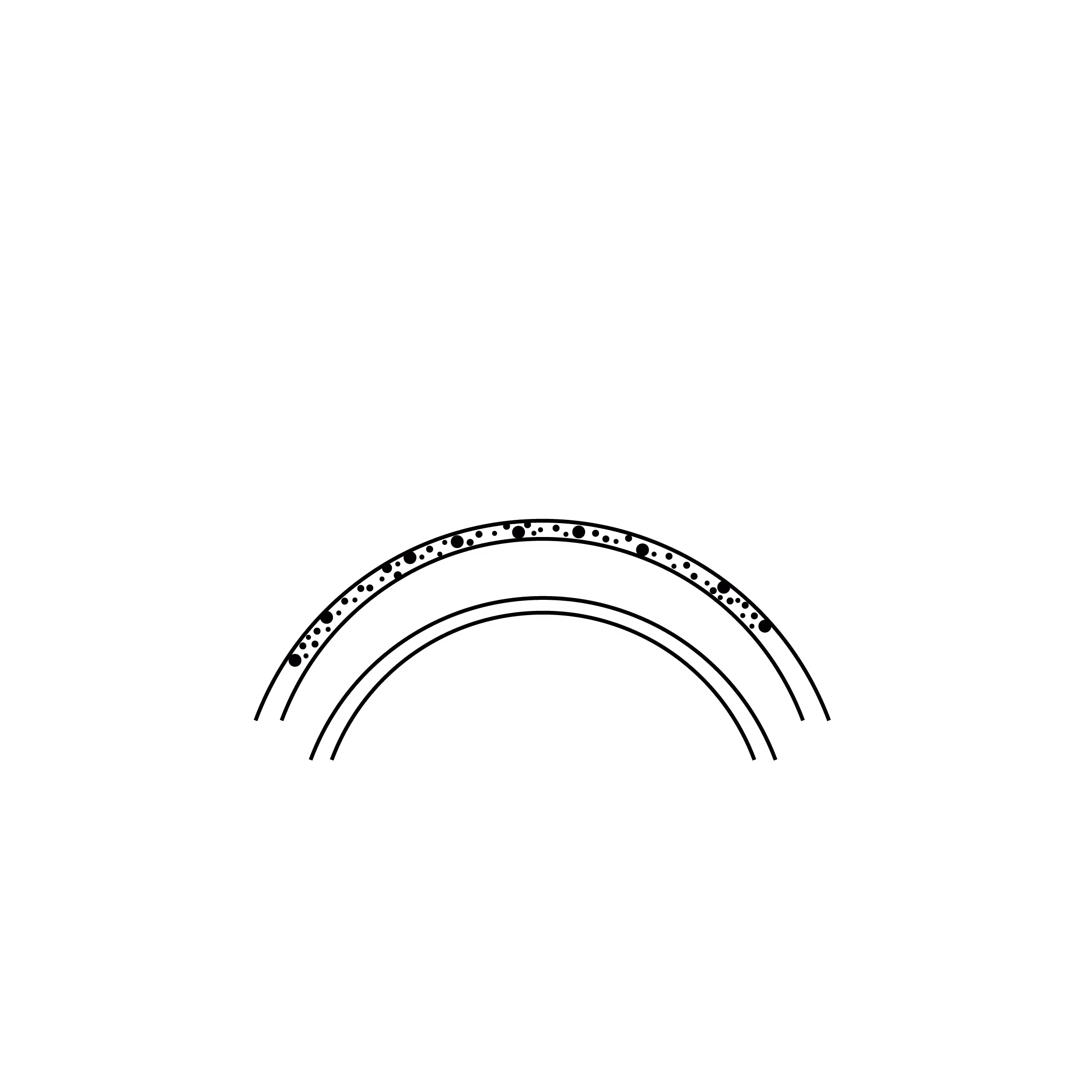
Epithelial and subepithelial dystrophies
Epithelial and subepithelial corneal dystrophies affect the superficial layers of the cornea. These dystrophies are characterised by abnormal deposits or cellular changes within or just below the epithelium (the outermost layer).
One of the most frequently encountered forms is Epithelial Basement Membrane Dystrophy (EBMD), also known as Map-Dot-Fingerprint Dystrophy or Cogan’s Dystrophy.
In this condition, the basement membrane, which acts as the "glue" for the surface cells, is abnormal. As a result, the epithelial cells fail to adhere correctly to the underlying layer, leading to the formation of microscopic bumps, folds, or thickened patches on the corneal surface.
These unstable areas often result in recurrent corneal erosions. Patients typically experience episodes of acute eye pain, particularly on waking, as the non-adherent surface cells are "torn" away by the eyelid when the eyes first open.
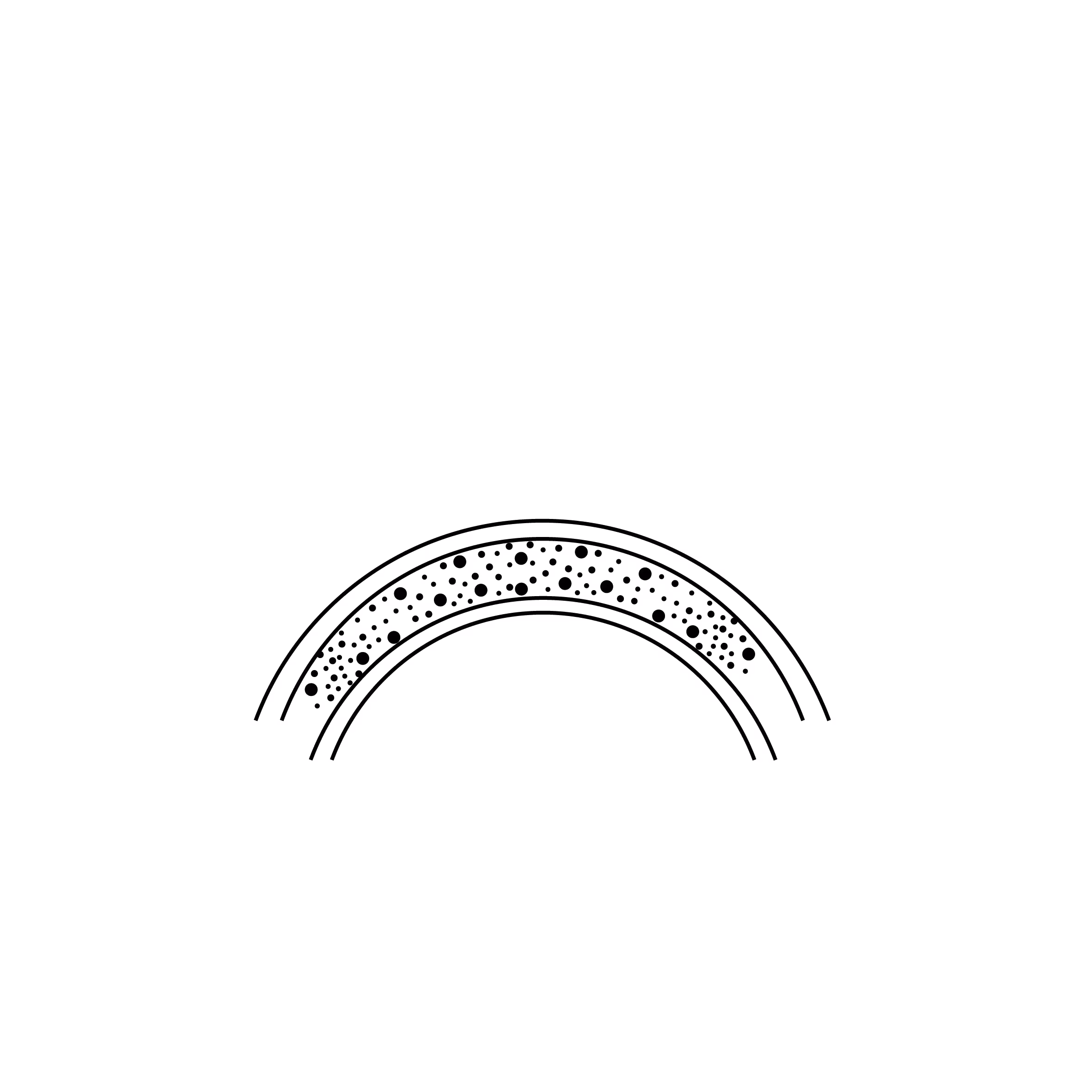
Stromal dystrophies
Epithelial-stromal corneal dystrophies can affect both the outer layer of the cornea (epithelium) and the deeper structural tissue beneath it (stroma). These dystrophies are characterised by the gradual accumulation of abnormal deposits within these layers, which can reduce corneal transparency and impair vision.
The clinical presentation varies depending on the specific type of dystrophy (such as Lattice, Granular, or Macular dystrophy), but common symptoms include:
- Blurred or hazy vision: As deposits accumulate, the cornea becomes less clear.
- Sensitivity to light (photophobia): Light scattering off the deposits can cause significant glare.
- Foreign body sensation: In some variants, the surface becomes irregular, leading to a persistent feeling of discomfort or grittiness.
- Visual distortion: Significant deposits can alter the shape of the cornea, leading to more pronounced disturbances in sight.
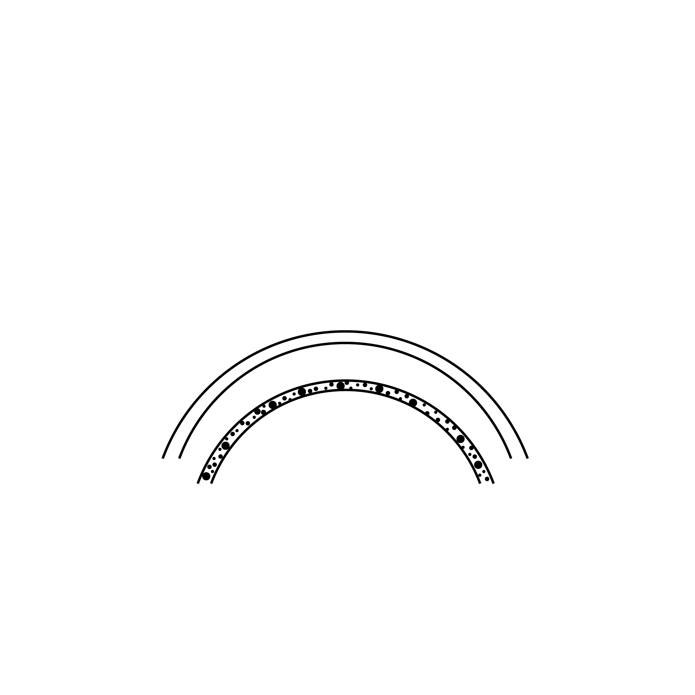
Endothelial dystrophies
Endothelial corneal dystrophies affect the endothelium, the innermost layer of the cornea. The endothelium acts as a vital pump, regulating fluid balance to ensure the cornea remains clear and transparent. In these dystrophies, the endothelial cells become dysfunctional, leading to fluid accumulation (oedema) and a loss of corneal clarity.
Symptoms often fluctuate throughout the day and may include:
- Blurred or misty vision: Typically most severe on waking, often improving as the day progresses and excess fluid evaporates.
- Visual aberrations: Halos around lights, glare and "starbursts" are common, particularly in low-light conditions.
- Pain and discomfort: In advanced cases, fluid-filled blisters can form on the corneal surface (bullous keratopathy). When these rupture, they cause acute pain and corneal erosions.
Fuchs’ endothelial dystrophy is one of the most prevalent forms of these conditions. In some regions, particularly in Northern Europe, it is estimated to affect up to 20% of the population.
Treatments
Treatment strategies are tailored to the specific type of dystrophy and the severity of the symptoms.
Epithelial and Stromal Dystrophies
Management focuses on maintaining a smooth ocular surface and restoring clarity:
- Artificial tears: Used regularly to alleviate irritation and maintain lubrication.
- "Bandage" contact lenses: These specialised lenses protect the corneal surface and promote healing.
- Surface Polishing (PTK/Trans-PTK): Laser procedures used to "smooth" the corneal surface and remove superficial opacities.
- Corneal Transplants (PKP, DALK): In cases of deep scarring or significant opacification, a transplant may be necessary to restore sight.
Endothelial Dystrophies
Treatment aims to manage fluid levels within the cornea:
- Hypertonic drops: These help to reduce oedema by drawing excess fluid out of the cornea.
- "Bandage" contact lenses: Used to provide relief from discomfort in cases of bullous keratopathy (fluid-filled blisters).
- Endothelial Lamellar Graft (e.g., DMEK): In advanced cases, surgery is often required. Modern techniques like DMEK replace only the damaged inner cell layer, leading to faster recovery and excellent visual outcomes.
Regular follow-up with an ophthalmologist is crucial to monitor the progression of the condition and ensure that treatment is adjusted at the appropriate time.
Motifs de consultations associés
Les procédures chirurchicales associées


Les questions fréquentes
Si vous avez d'autres questions n'hésitez pas à nous contacter !
Book a consultation
Swiss Visio Montchoisi
1006 Lausanne, Switzerland