Cataract surgery
Cataract surgery cataract is an ophthalmological procedure aimed at removing the eye's natural lens (crystalline lens) that has become clouded and replacing it with an intraocular lens (IOL) artificial transparent one. Generally performed under local/topical anesthesia, it is most often done by phacoemulsification, a minimally invasive technique that uses ultrasound to fragment and aspirate the lens. The procedure is quick, painless, and allows for rapid visual recovery within a few days.
Cataract surgery is a highly refined procedure that benefits from constant improvements, making it one of the most commonly performed and safest procedures worldwide.

How does it work?
Learn more about the procedure / equipment
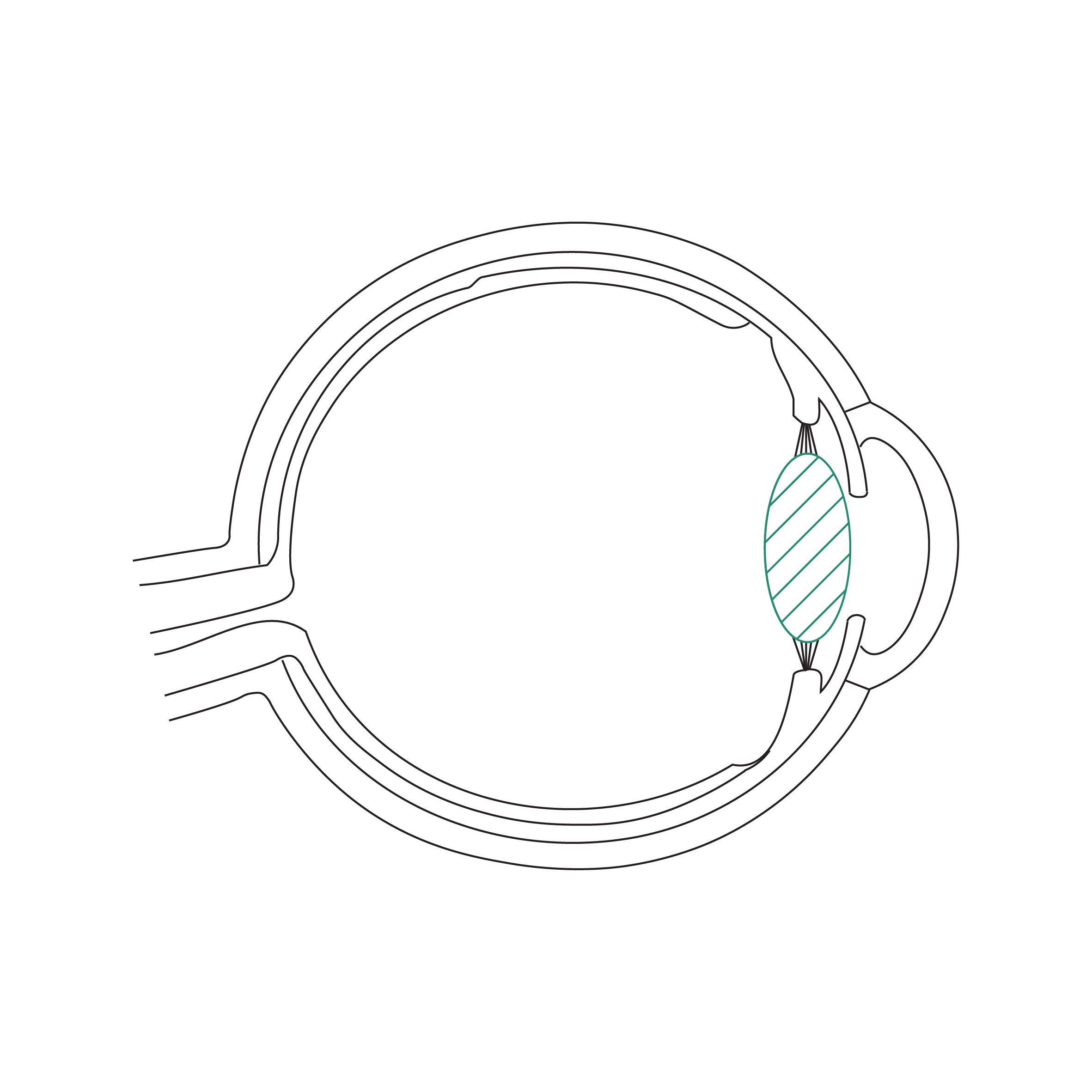
This illustration shows a complete eye in cross-section, viewed from the side, with an opacified lens at the front (the anterior segment), also known as a cataract.
The eye is shown in a horizontal position, as it would be when standing upright.

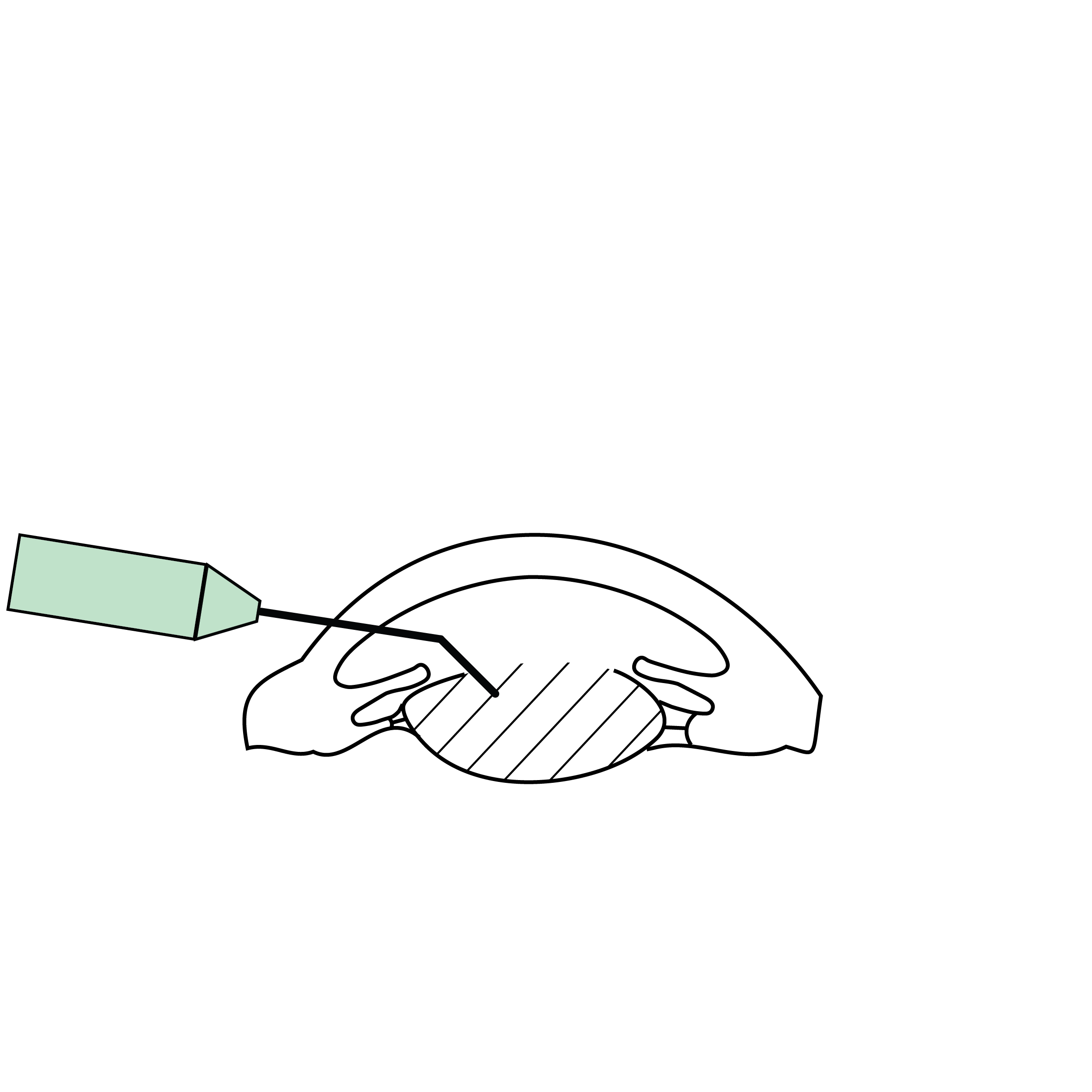
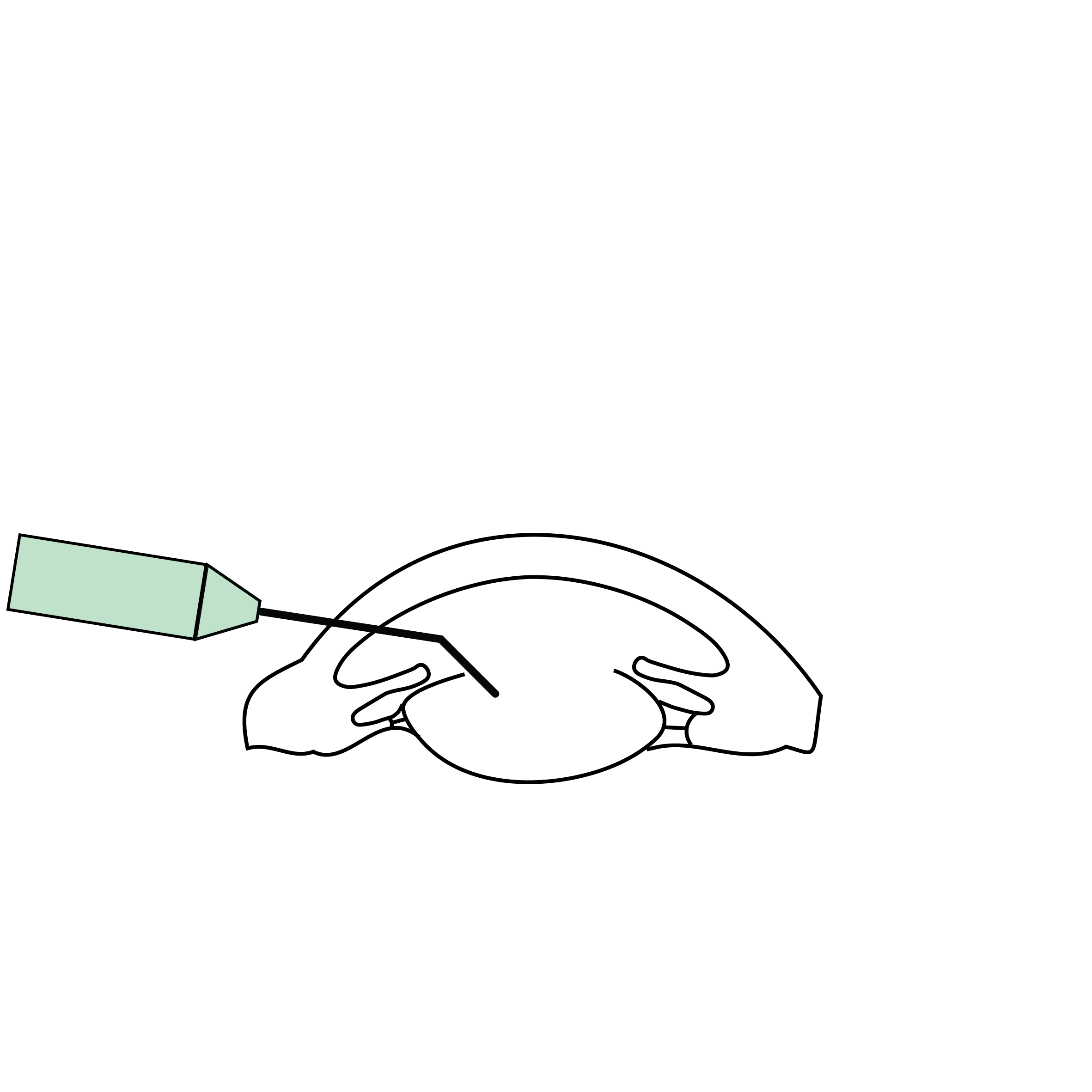
This drawing depicts only the anterior segment of the eye, viewed in profile cross-section. The eye is in a vertical position, as it would be when lying on one's back.
The eye's natural lens (the crystalline lens) has become cloudy, preventing light from entering the eye properly and thus affecting vision.
Cataract surgery: the cataract (a cloudy lens) is broken up into small pieces using ultrasound, a painless procedure.

Intraocular Lens (IOL): A new, clear artificial lens replaces the clouded natural lens to restore vision.
What is cataract surgery?
Cataract surgery is a common procedure that involves removing the eye's natural lens, also known as the crystalline lens, which has become opaque and cloudy, and replacing it with an artificial intraocular lens (IOL) (called a pseudophakic lens) to restore vision.
The procedure is generally performed under local or topical anesthesia (via eye drops), most often by phacoemulsification, a minimally invasive technique that uses ultrasound to fragment the lens before aspiration.
The procedure is quick and painless. It lasts approximately 10 to 15 minutes and is performed as an outpatient procedure, meaning you can go home the same day.
What is the preparation process?
During the first consultation, the ophthalmologist performs a comprehensive eye examination to confirm the cataract diagnosis and rule out other eye conditions that could affect the surgical outcome (e.g., glaucoma, age-related macular degeneration, etc.).
A series of precise eye measurements is taken to select the intraocular lens best suited to the patient's needs. These measurements include:
- Ocular biometry: measures, among other things, the axial length of the eye and the curvature of the cornea to calculate the power of the intraocular lens (IOL).
- Corneal topography: analyzes the shape and regularity of the cornea to determine eligibility for different types of IOLs.
- Astigmatism assessment: to determine if a toric IOL is needed.
- Specular microscopy: measures the density of corneal endothelial cells to determine the risk of persistent corneal edema after surgery.
- OCT of the posterior pole: confirms the condition of the macula and optic nerve to guide the choice of IOL to be used during surgery.
Choice of intraocular lens
Depending on the examination results and the patient's visual expectations, different lens options may be offered:
- Monofocal IOL: Clear vision without glasses for distance or near. Most patients choose clear distance vision and glasses for reading, but the opposite is also possible.
- Premium IOL: correction of distance, intermediate, and near vision without glasses.
- Toric IOLs: astigmatism correction.
The choice is discussed with the patient, taking into account their lifestyle and expectations.
How does recovery proceed after the procedure?
Visual recovery is rapid and progressive, depending on the degree of your cataract. Some patients regain clear vision as early as the day after the procedure, and most show significant improvement within a few days. Complete stabilization can sometimes take several weeks.
However, it is essential to carefully follow post-operative recommendations to ensure optimal healing and minimize the risk of complications. Here are the main steps and advice regarding the recovery period:
1. Immediately after the procedure
- The surgery is performed on an outpatient basis, allowing the patient to return home the same day.
- An eye shield is usually placed over the operated eye to prevent accidental trauma, especially during sleep.
- Vision may be blurry or hazy in the first few hours.
- A sensation of slight discomfort, stinging, or a foreign body is normal, but there should be no intense pain.
2. The first few days post-surgery
- Visual recovery begins the day after, with a progressive improvement in sharpness and color perception. Some patients already have clear vision.
- The patient must avoid rubbing the operated eye and keep the eye clean. Only post-operative eye drops should come into contact with the eye (no running water or other non-sterile liquids).
- Post-operative treatment is prescribed, generally including:
- Antibiotic eye drops to prevent infections.
- Anti-inflammatory eye drops (corticosteroids and/or NSAIDs) to accelerate recovery.
- Artificial tears and saline solution, optional, for cleaning the ocular surface.
- Intense physical activities, heavy lifting, and dusty environments should be avoided during the first week.
- Patients can generally resume light activities and driving as soon as vision is stable, subject to the ophthalmologist's authorization.
3. Medical Follow-up
- A post-operative consultation is usually scheduled within 24 hours of the procedure to check for healing and the absence of complications.
- Regular follow-up is conducted at one week, then at one month, to assess visual progress and adjust treatment if necessary.
- If both eyes need to be operated on, the second procedure is usually scheduled after the first eye has recovered, typically one to a few weeks later.
4. Medium-Term Progression (2nd to 4th post-operative week)
- Vision continues to improve gradually, although full recovery may take several weeks, depending on the complexity of the cataract and the overall condition of the eye.
- Adjustment of corrective glasses, if necessary, is usually done after one month, once vision is stable.
5. Long-Term Progression (months to years)
- Intraocular lenses (IOLs) are made from biocompatible materials designed to remain transparent for life so that you won't need to have them changed.
- The capsule that holds the IOL can thicken and become opaque (posterior capsular opacification = secondary cataract). This can occur several months or years after the procedure and be treated with a YAG laser capsulotomy. YAG laser.
Possible Risks and Complications
Although cataract surgery is a very safe procedure, some rare complications can occur, such as:
- Fragility of the capsule or its supporting fibers (zonules): requiring IOL implantation outside the natural lens capsule, which can be done during cataract surgery or in a subsequent procedure.
- Infection (endophthalmitis): approximately 0.08% risk, manifesting as intense pain, significant redness, and sudden vision loss, requiring emergency treatment.
- Corneal or macular edema: delaying visual recovery.
- Retinal detachment: more common in patients with high myopia.
Long-term results
Most patients regain clear and stable vision, allowing them to resume their daily activities without issue.
Some may require glasses for reading or certain precision activities, depending on the type of artificial intraocular lens (IOL) implanted.
Regular follow-up with an ophthalmologist is recommended to monitor eye health, especially for patients with other conditions such as glaucoma or age-related macular degeneration.
By following recommendations and undergoing rigorous follow-up, recovery after cataract surgery is generally very satisfactory, leading to a significant improvement in quality of life.
What are the options for vision adjustment after surgery?
Add-on (or piggyback) IOL implantation:
- A secondary intraocular lens is added in front of the existing IOL, in the ciliary sulcus, to refine the correction.
- This solution is reversible and allows for precise refractive adjustment (myopia, hyperopia, astigmatism, presbyopia).
- It is particularly indicated in cases of significant refractive errors or an evolving need for correction.
Laser correction:
- When the IOL is in place and further adjustment is needed, laser corneal refractive surgery (LASIK, SMILE, PRK) is effective.
- Used to correct mild to moderate residual refractive errors (myopia, farsightedness or astigmatism).
- Suitable for patients wishing to reduce their dependence on glasses after surgery.
- A quick, minimally invasive procedure with a short recovery time.
IOL Explantation and Replacement:
- In cases of significant refractive error or intolerance to the initial lens, the IOL can be removed and replaced with a more suitable lens.
- This is a more invasive procedure, generally considered as a last resort.
Les différentes types de greffes
Découvrez les kératoplasties (= greffes de cornée)
Frequently asked questions
If you have any further questions, please do not hesitate to contact us!
When should I consider cataract surgery?
When your vision becomes blurry, you experience difficulty with daily activities (such as reading or driving), or glasses/contact lenses can no longer improve your vision.
Is cataract surgery painful?
No, the procedure is usually performed under local or topical (eye drop) anesthesia, and you will not feel any pain during the operation. If necessary, sedation or general anesthesia may be used.
How long does cataract surgery last?
Typically, the procedure only takes about ten minutes, and you can go home the same day.
When will my vision return to normal after cataract surgery?
Vision improves within the first few days, sometimes becoming clear as early as the day after the operation. Full recovery can take several weeks.
Will I need to wear glasses after cataract surgery?
This depends on the type of intraocular lens (IOL) implanted. Glasses may be necessary for reading or certain activities. Premium IOLs reduce the need for glasses.
What is the lifespan of the intraocular lens?
The lens is designed to last a lifetime and generally does not require replacement.
Can I resume my normal activities quickly after cataract surgery?
Most light activities can be resumed after a few days, but intense physical exertion should be avoided for at least one week.
Do I need to take any special precautions after cataract surgery?
Yes, keep your eye clean, avoid rubbing it, exposure to dust, swimming, and follow the prescribed treatment.
Are there any dietary restrictions after cataract surgery?
No, no specific dietary restrictions are necessary.
Can I drive after cataract surgery?
Wait for your ophthalmologist's advice, usually within the first few days post-surgery if your vision is stable and sufficient.
Is it possible to operate on both eyes for cataracts at the same time?
Typically, each eye is operated on separately, with an interval of one to several weeks between procedures. If necessary or if you prefer, it is possible to operate on both eyes on the same day, in consultation with your surgeon.
Does cataract surgery improve all eye conditions?
No, it only corrects cataracts. If other eye diseases are present (e.g., glaucoma, macular degeneration), they require specific monitoring and may limit the final visual acuity.
Is there an age limit for cataract surgery?
No, age is not a contraindication as long as there is a cataract significantly impacting your vision.
Can the eye reject the intraocular lens?
No, the lens is biocompatible and does not cause rejection.
What to do in case of pain or sudden vision loss after cataract surgery?
Consult your ophthalmologist immediately or go to the nearest ophthalmic emergency center, as this could be a sign of a complication requiring prompt attention.
Linked procedures
Discover other related surgical topics


Linked conditions


Book a consultation
Swiss Visio Montchoisi
1006 Lausanne, Switzerland