Keratoconus: how the disease progresses and when to intervene
Keratoconus is not managed the same way depending on whether it is progressing or has settled. Measured progression is what drives the decision.
Created on
27.09.2026
·
Updated on
11.10.2026

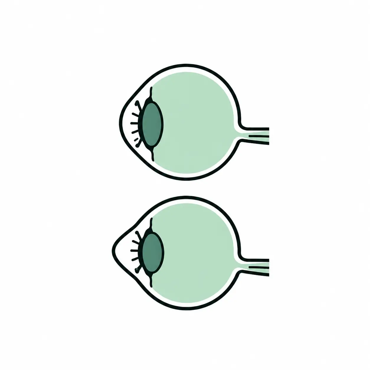
Keratoconus is a progressive corneal disease, and it is that progression, measured from one examination to the next, that determines treatment. Stable keratoconus is monitored. Progressive keratoconus is stabilised. A cornea that is already badly distorted calls for different procedures altogether. This article sets out how the disease advances, how that advance is measured, and at what point each option is discussed at the consultation.
A disease judged on its trajectory, not on a snapshot
Keratoconus gradually thins and distorts the cornea, the transparent lens at the front of the eye, which takes on a cone shape instead of remaining evenly curved. That distortion creates an irregular astigmatism that ordinary spectacles cannot fully correct.
The condition usually affects both eyes, sometimes asymmetrically. It frequently begins between the ages of 15 and 25, then tends to stabilise after 35 to 40. According to the European Society of Cataract and Refractive Surgeons (ESCRS), it affects around 1.2 people per 1,000.
Two patients with the same degree of distortion can therefore warrant opposite decisions: in one, the cornea is still changing and needs stabilising; in the other, it has not moved for years and the question becomes purely optical. This is why keratoconus is never managed on the strength of a single examination.
How progression is measured
Progression in keratoconus is a documented change in the shape and thickness of the cornea between two examinations. It is not inferred from an impression, it is mapped.
The key examinations
Corneal topography and tomography map the surfaces and the various layers of the cornea, and detect very early distortion before any drop in vision. They are complemented by optical coherence tomography (OCT) of the cornea and by pachymetry, the measurement of corneal thickness. Together these establish the diagnosis, assess the stage and track the course of the disease.
What is compared from one visit to the next
- any increase in the distortion and any shift in its apex
- corneal thickness, particularly at the thinnest point
- any worsening of the irregular astigmatism
- the optical correction required, and what it actually achieves
- contact lens tolerance, often the first functional sign of a change
Needing frequent changes of prescription, with astigmatism on the increase, justifies a corneal examination. The early signs of keratoconus are covered in a dedicated article.
At what stage do you intervene?
The options follow from two questions: is the cornea still changing shape, and is vision still correctable? The first governs stabilisation, the second optical improvement. These are distinct aims and are often combined.
Early, stable keratoconus: monitoring
Stable keratoconus does not always call for immediate treatment. Management combines regular monitoring by topography with a suitable optical correction. Spectacles suffice while the astigmatism remains regular; rigid contact lenses take over once it becomes too irregular.
Progressive keratoconus: stabilising the cornea
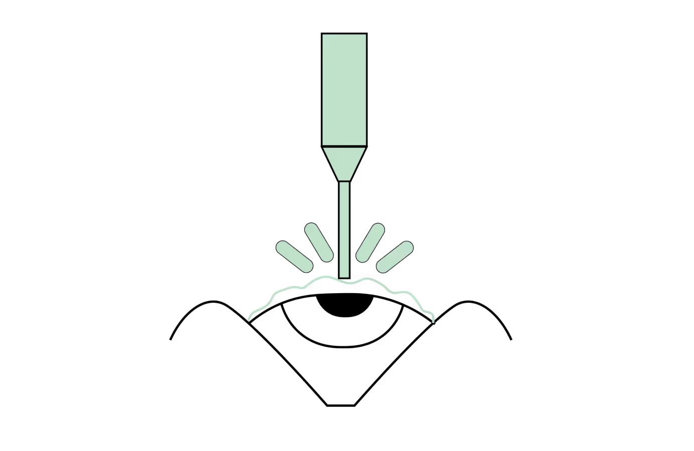
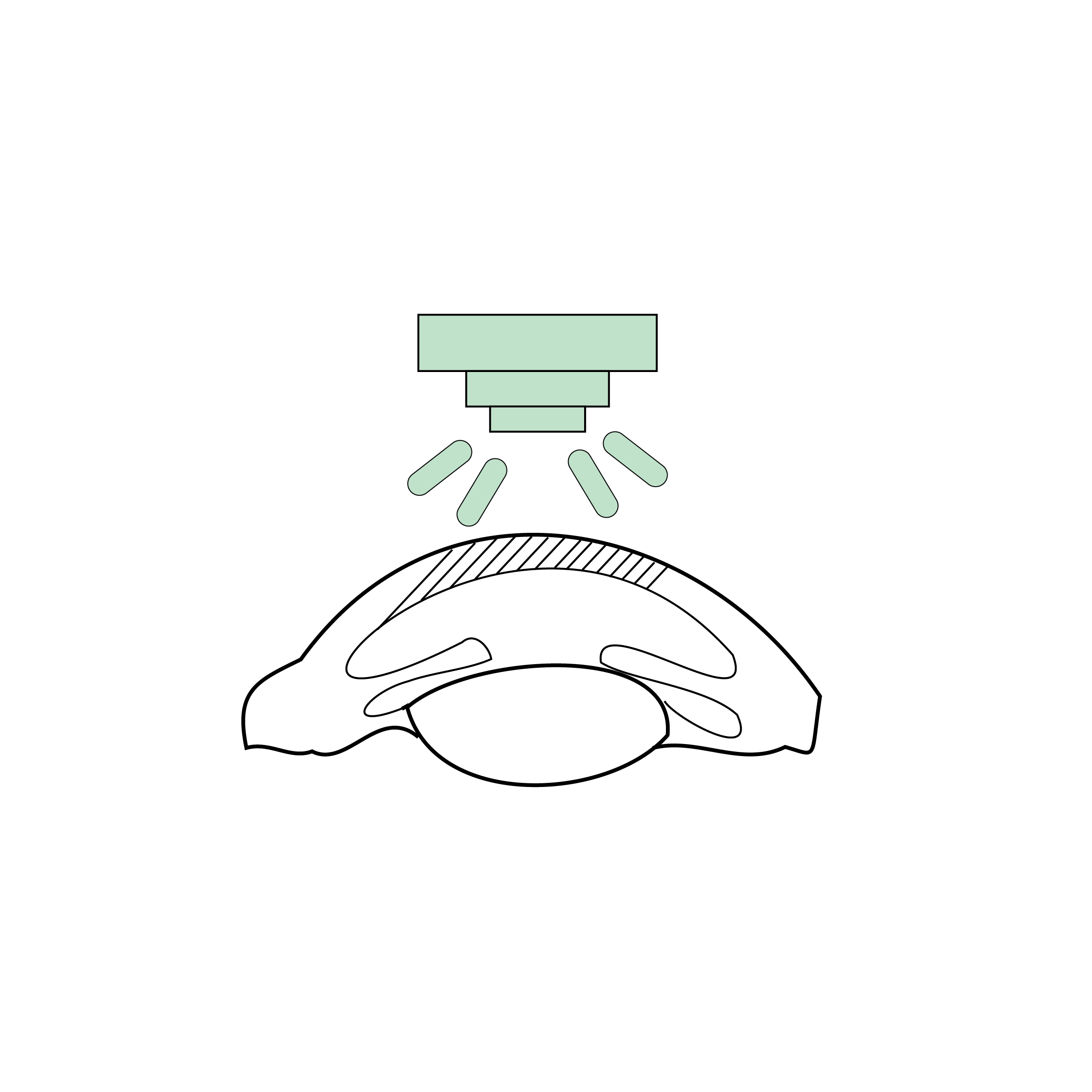
As soon as progression is documented, the objective changes: the distortion has to be stopped. Corneal collagen cross-linking (CXL) is the reference treatment for progressive keratoconus. It creates new bonds between the collagen fibres to stiffen the cornea, by applying riboflavin and then delivering a controlled dose of UVA light.
Adolescents and young adults are the first concerned, since the disease advances quickly at that age. The course of a cross-linking procedure, its aftermath and its limits are covered separately. The essential point here: cross-linking stabilises, it does not restore vision already lost.
Established distortion: improving shape and vision
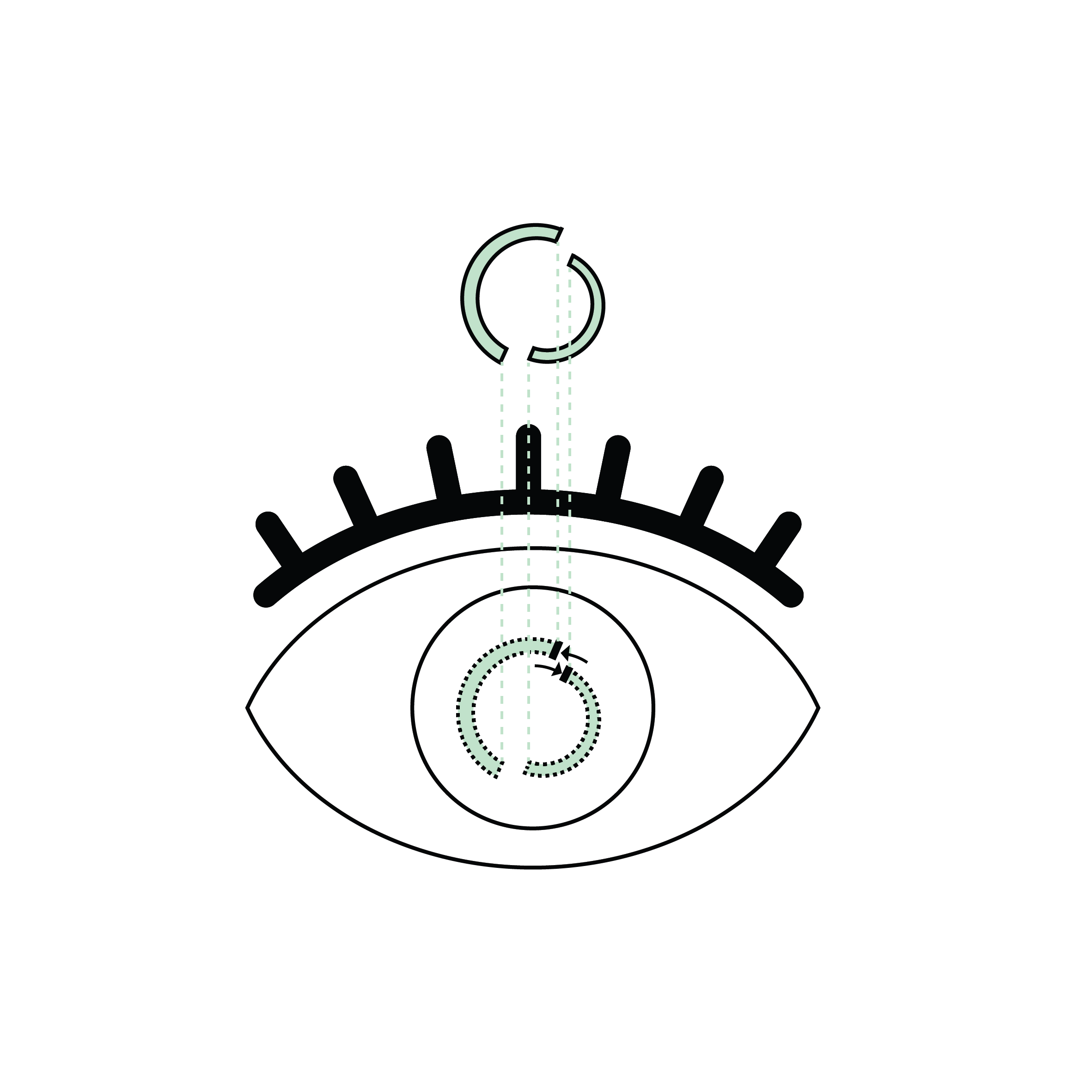
Where the distortion is already marked and optical correction no longer suffices, other procedures aim to regularise the cornea. Intracorneal rings and segments (ICRS and CAIRS) are inserted within the thickness of the cornea to reduce its distortion. A customised PRK guided by topography or by aberrations may also be discussed, sometimes alongside cross-linking.
These procedures address specific situations and are not suitable for every cornea. The indications for intracorneal rings are set out in a dedicated article.
Advanced stage: corneal transplantation
Corneal transplantation comes into play where distortion or clouding leaves no correction satisfactory. It is not an inevitable stage of keratoconus but an option of last resort. Depending on which layers are affected, deep anterior lamellar keratoplasty (DALK) replaces only the diseased portion while preserving the patient's own endothelium, whereas penetrating keratoplasty (PKP) replaces the full thickness of the cornea.
This table summarises the broad directions discussed at the consultation. It does not replace an assessment and does not prejudge any individual indication.
What tips the balance from monitoring to treatment
The move from monitoring to treatment does not rest on a single threshold. Several factors weigh together in the decision:
- documented progression between two examinations, the principal argument
- the patient's age, the disease advancing faster in adolescents and young adults
- whether the second eye is affected, and how it is behaving
- the vision actually achieved with the best available correction
- the presence of active aggravating factors, such as eye rubbing or untreated ocular allergy
Conversely, a cornea stable for several years in a 40-year-old patient, comfortably corrected, does not necessarily warrant a procedure. Keratoconus surgery only makes sense in relation to a trajectory.
The one lever that stays in your hands
Chronic eye rubbing is a major aggravating factor in keratoconus: it applies repeated mechanical stress to an already weakened cornea. It often goes with ocular allergy or eczema, which keep the itching going.
Treating the allergy and breaking the habit of rubbing are therefore an integral part of management. A family history of keratoconus also justifies a corneal examination in close relatives, even without symptoms, since topography detects distortion before vision drops.
The two eyes do not progress at the same pace
Keratoconus usually affects both eyes, but sometimes markedly asymmetrically. It is not unusual for one eye to be well distorted while the other still looks more or less normal. That asymmetry explains two situations seen often at the consultation.
The first: a patient attends only for the eye that bothers them, when the second deserves the same mapping. Early distortion is frequently detectable there on topography well before vision drops. The second: the two eyes may warrant different decisions at the same time, cross-linking on one side and simple monitoring on the other. Follow-up therefore always covers both eyes, whatever the symptoms.
What keratoconus does not mean
Several fears come up regularly and deserve to be laid to rest. Keratoconus does not lead to blindness: it degrades the quality of vision through irregular astigmatism, and the treatments available today preserve functional vision in the great majority of cases.
A diagnosis of keratoconus does not call for immediate surgery either. Many patients are simply monitored for years. Nor does keratoconus condemn anyone to rigid contact lenses for life: regularising the cornea sometimes allows a return to a simpler correction. Equally, keratoconus diagnosed early should not be dismissed as harmless because vision is still good, since that is precisely the window in which stabilisation preserves the most.
In practice at Dr Hammer's clinic
Every keratoconus plan begins with a full corneal assessment: topography and tomography, corneal OCT, pachymetry, and comparison with earlier examinations where they exist. It is that comparison which separates stable from progressive keratoconus, and which shapes the whole decision.
Dr Arthur Hammer is an FMH ophthalmic surgeon, MD-PhD, FEBO and FEBOS-CR, practising in Lausanne and Geneva. Keratoconus surgery is among his areas of practice, from cross-linking through to lamellar transplants. His background and areas of expertise are set out on the site.
If you are looking for a specialist opinion, the page on keratoconus treatment in Switzerland sets out how consultations work in Lausanne and Geneva.
When to seek advice without delay
Some signs point to rapid progression and warrant prompt ophthalmic advice:
- a drop in vision over a few weeks
- distortion or double images that are becoming more marked
- astigmatism changed several times in a short period
- growing intolerance of contact lenses
- pain or unusual redness in a known keratoconus
These symptoms may suggest progression, but only a specialist examination with topography can confirm it.
FAQ: common questions about keratoconus progression
At what age does keratoconus stop progressing?
Keratoconus frequently begins between the ages of 15 and 25, then tends to stabilise after 35 to 40. That general tendency does not remove the need for individual monitoring: some cases continue to progress beyond that, others settle far earlier. Only the comparison of corneal topographies over time can establish whether a given patient's cornea is still changing shape.
How do you know that keratoconus is progressing?
Progression is documented by comparing several corneal topographies and tomographies taken at regular intervals. What is examined is the change in the distortion, in corneal thickness and in the irregular astigmatism. Frequent changes of prescription are a warning sign, but they do not replace those examinations: it is the mapping of the cornea that settles the question.
Does stable keratoconus need treatment?
Stable keratoconus does not always call for immediate cross-linking. Regular monitoring may be enough, combined with a suitable optical correction: spectacles first, then rigid contact lenses once the astigmatism becomes too irregular. The decision to treat rests on documented progression, not on the diagnosis alone.
Does cross-linking restore vision that has already been lost?
No. The aim of cross-linking is to halt the progression of keratoconus, not to restore vision that has already deteriorated. A slight improvement in corneal shape is sometimes observed, but it is not guaranteed and is not the purpose of the treatment. That is precisely why intervening early matters: the less distorted the cornea when it is stabilised, the better the vision preserved.
Can you have LASIK if you have keratoconus?
Established keratoconus rules out conventional laser refractive surgery, which would further weaken an already distorted cornea. Suspected keratoconus found on topography calls for the same caution. Certain customised surface techniques, guided by aberrations, may however be discussed once the cornea is stable, within a carefully defined setting and on the surgeon's indication.
Does keratoconus always end in a corneal transplant?
No, and this is a common worry at the consultation. Transplantation concerns keratoconus that has reached an advanced stage, where distortion or clouding prevents any satisfactory correction. Stabilising the cornea early reduces the likelihood of reaching that point. Where a transplant is indicated, lamellar techniques often allow only the affected layers to be replaced.
Is cross-linking covered in Switzerland?
Where it treats documented progressive keratoconus, cross-linking is in principle covered by Swiss basic insurance (LAMal), subject to the deductible and the 10% co-payment. The exact terms depend on the situation and are set out at the consultation. That cover assumes established progression, which is why follow-up examinations matter so much.
Does stopping rubbing your eyes really make a difference?
Yes. Chronic eye rubbing is a major aggravating factor in keratoconus: it applies repeated mechanical stress to an already weakened cornea. Treating the allergy or eczema that triggers the urge to rub is therefore part of management, on the same footing as any surgical step. It is also the one lever entirely in the patient's hands.
In summary
Keratoconus is managed according to its trajectory, not its appearance at a single moment. While the cornea is still changing shape, the aim is to stabilise it, and cross-linking is the reference treatment for that. Once the cornea is stable, the question becomes optical and other options come into play. Transplantation remains a fallback, not a foregone conclusion.
Documenting progression takes repeated examinations over time: that is the starting point of every decision. For a corneal assessment or a second opinion, you can book an appointment with Dr Arthur Hammer, FMH ophthalmologist specialising in corneal and refractive surgery in Lausanne and Geneva.



Linked procedures
.avif)
.avif)


Related articles
Discover our other articles that might interest you

Book a consultation
Swiss Visio Montchoisi
1006 Lausanne, Switzerland